Difference between revisions of "Small Animal Dermatology Q&A 05"
Ggaitskell (talk | contribs) (Created page with "{{Template:Manson Moriello}} centre|500px<br>centre|500px <br /> '''An 8-month-old...") |
Ggaitskell (talk | contribs) |
||
| Line 13: | Line 13: | ||
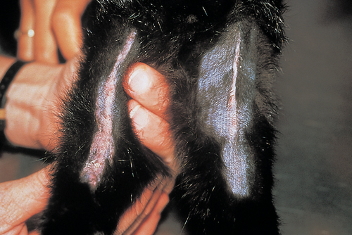
This is a classic presentation of feline eosinophilic granuloma. <br><br> | This is a classic presentation of feline eosinophilic granuloma. <br><br> | ||
Other presentations include firm swellings on the chin (fat chin syndrome), swollen lower lips (pouting cat syndrome), oral lesions in the mouth, firm nodules in the skin, and nodules on the ear pinnae. | Other presentations include firm swellings on the chin (fat chin syndrome), swollen lower lips (pouting cat syndrome), oral lesions in the mouth, firm nodules in the skin, and nodules on the ear pinnae. | ||
| − | |l1= | + | |l1=Feline Eosinophilic Granuloma |
|q2=What are the treatment options? | |q2=What are the treatment options? | ||
|a2= | |a2= | ||
| Line 19: | Line 19: | ||
*Methylprednisone acetate (20 mg/cat SC), every 2 weeks until the lesions resolve (4–6 weeks), is effective.<br><br> | *Methylprednisone acetate (20 mg/cat SC), every 2 weeks until the lesions resolve (4–6 weeks), is effective.<br><br> | ||
*Recurrent lesions suggest an underlying trigger such as FAD, food allergy, and/or atopy. | *Recurrent lesions suggest an underlying trigger such as FAD, food allergy, and/or atopy. | ||
| − | |l2= | + | |l2=Steroids |
|q3=What components of eosinophil granules may be responsible for collagen degradation? | |q3=What components of eosinophil granules may be responsible for collagen degradation? | ||
|a3= | |a3= | ||
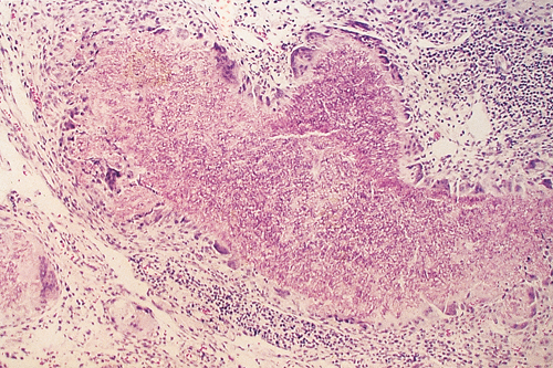
The pathogenesis of these lesions is unknown. However, tissue damage may be caused by eosinophil collagenase that degrades type I and II collagen and gelatinases that degrade type XVII collagen. | The pathogenesis of these lesions is unknown. However, tissue damage may be caused by eosinophil collagenase that degrades type I and II collagen and gelatinases that degrade type XVII collagen. | ||
| − | |l3= | + | |l3=Eosinophils |
</FlashCard> | </FlashCard> | ||
Revision as of 17:51, 6 June 2011
| This question was provided by Manson Publishing as part of the OVAL Project. See more small animal dermatological questions |
An 8-month-old kitten is presented for raised, firm, pencil-like lesions on the caudal aspects of both hind legs. The owner reports the lesions developed rapidly but do not seem bothersome to the kitten. Dermatological examination reveals hard, linear lesions in the superficial dermis. Skin biopsies reveal eosinophilic granulomatous inflammation and collagen degeneration.
| Question | Answer | Article | |
| What is the diagnosis and what are other clinical presentations of the same ‘syndrome’? | This is a classic presentation of feline eosinophilic granuloma. |
Link to Article | |
| What are the treatment options? |
|
Link to Article | |
| What components of eosinophil granules may be responsible for collagen degradation? | The pathogenesis of these lesions is unknown. However, tissue damage may be caused by eosinophil collagenase that degrades type I and II collagen and gelatinases that degrade type XVII collagen. |
Link to Article | |

