Difference between revisions of "Avian Medicine Q&A 21"
| Line 15: | Line 15: | ||
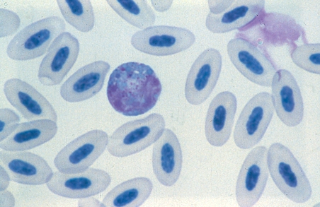
The heterophil shows increased cytoplasmic basophilia, abnormal cytoplasmic granules that include round, basophilic primary granules. This would be considered a 2+ to 3+ toxicity. | The heterophil shows increased cytoplasmic basophilia, abnormal cytoplasmic granules that include round, basophilic primary granules. This would be considered a 2+ to 3+ toxicity. | ||
| − | |l1= | + | |l1= |
|q2= What is the significance of the cell demonstrated in the figure? | |q2= What is the significance of the cell demonstrated in the figure? | ||
|a2= Toxic changes in the heterophils represent the presence of a septicaemic or toxaemic condition, e.g. bacterial toxins. | |a2= Toxic changes in the heterophils represent the presence of a septicaemic or toxaemic condition, e.g. bacterial toxins. | ||
| Line 22: | Line 22: | ||
Therefore, the presence of numerous heterophils showing signs of marked toxicity represents a severe condition and hence a poor prognosis for survival. | Therefore, the presence of numerous heterophils showing signs of marked toxicity represents a severe condition and hence a poor prognosis for survival. | ||
| − | |l2= | + | |l2= |
</FlashCard> | </FlashCard> | ||
| Line 29: | Line 29: | ||
desc none}} | desc none}} | ||
[[Category: Avian Medicine Q&A]] | [[Category: Avian Medicine Q&A]] | ||
| + | [[Category:To Do - Manson]] | ||
Revision as of 21:34, 2 August 2011
| This question was provided by Manson Publishing as part of the OVAL Project. See more Avian Medicine questions |
A mitred conure (Psittacara mitrata) was presented with a 3 day history of weakness, lethargy and anorexia. The physical examination revealed a moderate reduction of the pectoral muscle mass and slight dyspnoea. The bird was weak, but able to perch. A blood profile revealed a mild to moderate leucocytosis at 18.6 × 10⁹ l-1, heterophilia at 17 × 109 l-1 and lymphopenia at 0.93 × 10⁹ l-1.
| Question | Answer | Article | |
| What is the cell demonstrated in the Wright’s-stained peripheral blood film above? | The slide demonstrates a toxic heterophil.
The heterophil shows increased cytoplasmic basophilia, abnormal cytoplasmic granules that include round, basophilic primary granules. This would be considered a 2+ to 3+ toxicity. |
[[|Link to Article]] | |
| What is the significance of the cell demonstrated in the figure? | Toxic changes in the heterophils represent the presence of a septicaemic or toxaemic condition, e.g. bacterial toxins.
The greater the degree of toxicity and number of cells affected, the more severe the condition. Therefore, the presence of numerous heterophils showing signs of marked toxicity represents a severe condition and hence a poor prognosis for survival. |
[[|Link to Article]] | |
