Difference between revisions of "Small Animal Orthopaedics Q&A 11"
Ggaitskell (talk | contribs) |
|||
| (One intermediate revision by the same user not shown) | |||
| Line 1: | Line 1: | ||
| − | |||
| − | |||
| − | |||
[[File:SmAnOrth 11.jpg|centre|500px]] | [[File:SmAnOrth 11.jpg|centre|500px]] | ||
| Line 14: | Line 11: | ||
|a1= | |a1= | ||
The dog has a retained cartilage core (RCC) associated with the distal ulnar physis and possibly retarded ulnar growth. | The dog has a retained cartilage core (RCC) associated with the distal ulnar physis and possibly retarded ulnar growth. | ||
| − | |l1= | + | |l1= |
|q2=What is the pathogenesis of this condition? | |q2=What is the pathogenesis of this condition? | ||
|a2= | |a2= | ||
| Line 26: | Line 23: | ||
While the distal ulnar physis is most commonly affected, RCCs have also been reported in the distal femur and distal tibia. | While the distal ulnar physis is most commonly affected, RCCs have also been reported in the distal femur and distal tibia. | ||
| − | |l2= | + | |l2= |
|q3=Which breeds of dogs are most commonly affected? | |q3=Which breeds of dogs are most commonly affected? | ||
|a3= | |a3= | ||
| Line 32: | Line 29: | ||
RCC is also reported in other species, including chickens, pigs and, possibly, horses. | RCC is also reported in other species, including chickens, pigs and, possibly, horses. | ||
| − | |l3= | + | |l3= |
</FlashCard> | </FlashCard> | ||
Revision as of 08:41, 2 September 2011
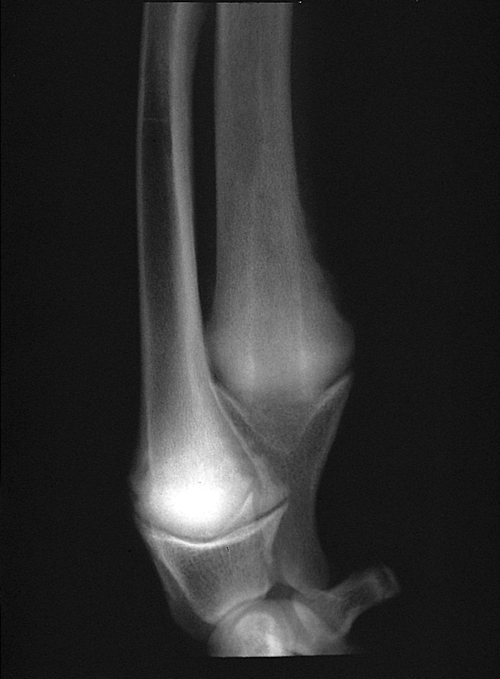
A lateral view radiograph of the left distal antebrachium of a five-month-old, male Irish Wolfhound with a history of a left forelimb lameness and mild valgus deformity of the left carpus.
| Question | Answer | Article | |
| What is the radiographic diagnosis? | The dog has a retained cartilage core (RCC) associated with the distal ulnar physis and possibly retarded ulnar growth. |
[[|Link to Article]] | |
| What is the pathogenesis of this condition? | RCCs (retained cartilage cores, retained endochondral cartilage cores) are caused by retarded endochondral ossification. RCCs are often bilateral, often clinically silent and may resolve uneventfully. The characteristic lesion is a cone-shaped mass of unmineralized hypertrophic cartilage with its base located at the center of the distal ulnar growth plate and its apex projecting into the metaphysis. The periphery of the physis is normal. The cause of the abnormality is unknown. One therory is that an interruption of oxygenation to a portion of the physis, perhaps due to micro trauma, results in an inability of the local environment to transform the cartilage into bone. While the distal ulnar physis is most commonly affected, RCCs have also been reported in the distal femur and distal tibia. |
[[|Link to Article]] | |
| Which breeds of dogs are most commonly affected? | Great Danes and Irish Wolfhounds. RCCs have also been reported in the Afghan, Collie and McKenzie Wolf. RCC is also reported in other species, including chickens, pigs and, possibly, horses. |
[[|Link to Article]] | |
