Difference between revisions of "Small Animal Abdominal and Metabolic Disorders Q&A 11"
Ggaitskell (talk | contribs) |
|||
| (One intermediate revision by the same user not shown) | |||
| Line 1: | Line 1: | ||
| − | |||
| − | |||
| − | |||
[[File:SmAn Ab and Met 11.jpg|centre|500px]] | [[File:SmAn Ab and Met 11.jpg|centre|500px]] | ||
| Line 18: | Line 15: | ||
The presence of erythrocytes indicates that some uterine tissue remains and makes the diagnosis simpler. | The presence of erythrocytes indicates that some uterine tissue remains and makes the diagnosis simpler. | ||
| − | |l1= | + | |l1= |
|q2=What techniques could be used to confirm this diagnosis? | |q2=What techniques could be used to confirm this diagnosis? | ||
|a2= | |a2= | ||
| Line 28: | Line 25: | ||
Investigative techniques include diagnostic laparoscopy or laparotomy. | Investigative techniques include diagnostic laparoscopy or laparotomy. | ||
| − | |l2= | + | |l2= |
|q3=What are the differential diagnoses for this condition? | |q3=What are the differential diagnoses for this condition? | ||
|a3= | |a3= | ||
| Line 41: | Line 38: | ||
Differential diagnoses for attractiveness to males includes vaginitis and anal gland disease. | Differential diagnoses for attractiveness to males includes vaginitis and anal gland disease. | ||
| − | |l3= | + | |l3= |
|q4=How should the condition be treated? | |q4=How should the condition be treated? | ||
|a4= | |a4= | ||
| Line 47: | Line 44: | ||
Frequently the remnant is associated with the right ovarian pedicle, and surgery is best performed during or shortly after oestrus when the ovarian tissue reaches its maximum size. | Frequently the remnant is associated with the right ovarian pedicle, and surgery is best performed during or shortly after oestrus when the ovarian tissue reaches its maximum size. | ||
| − | |l4= | + | |l4= |
</FlashCard> | </FlashCard> | ||
Revision as of 09:50, 2 September 2011
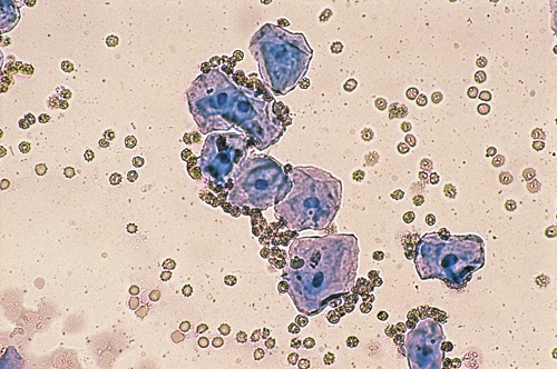
A three-year-old, female Collie-cross had a routine ovariohysterectomy at 18 months of age. Since that time she has had clinical signs of oestrus approximately every seven months and has been attractive to male dogs. Presently she has a red-tinged vulval discharge. A vaginal smear has been obtained and stained with a modified Wright-Giemsa stain.
| Question | Answer | Article | |
| What are the features of the vaginal smear, and what is the likely diagnosis? | Both large and small intermediate epithelial cells and some erythrocytes are present within the smear. These changes are associated with an elevation in plasma oestrogen concentration, and the likely diagnosis is that of ovarian remnant syndrome. The presence of erythrocytes indicates that some uterine tissue remains and makes the diagnosis simpler. |
[[|Link to Article]] | |
| What techniques could be used to confirm this diagnosis? | Collection of a vaginal smear when the bitch has clinical signs of oestrus provides a simple and rapid method of diagnosis. It is also possible to measure peripheral plasma progesterone concentrations two weeks after the signs of oestrus have disappeared. Raised progesterone concentrations are diagnostic for luteal tissue present within the ovarian remnant. Investigative techniques include diagnostic laparoscopy or laparotomy. |
[[|Link to Article]] | |
| What are the differential diagnoses for this condition? | The differential diagnoses for recurrent vulval discharge in an ovariohysterectomized bitch include:
Differential diagnoses for attractiveness to males includes vaginitis and anal gland disease. |
[[|Link to Article]] | |
| How should the condition be treated? | Surgical removal of the ovarian remnant will be curative. Frequently the remnant is associated with the right ovarian pedicle, and surgery is best performed during or shortly after oestrus when the ovarian tissue reaches its maximum size. |
[[|Link to Article]] | |
