Difference between revisions of "Cytology Q&A 17"
Ggaitskell (talk | contribs) (Created page with "{{Template:Manson}} [[Image:|centre|500px]] <br /> '''A seven-year-old neutered male Jack Russell Terrier presented with an interdigital mass of several weeks’ duration. Sm...") |
Ggaitskell (talk | contribs) |
||
| (3 intermediate revisions by 3 users not shown) | |||
| Line 1: | Line 1: | ||
| − | {{Template:Manson}} | + | {{Template:Manson |
| + | |book = Cytology Q&A}} | ||
| − | [[Image:|centre|500px]] | + | [[Image:Cytology 17a.JPG|centre|500px]] |
| + | <br> | ||
| + | [[Image:Cytology 17b.JPG|centre|500px]] | ||
<br /> | <br /> | ||
| Line 17: | Line 20: | ||
*The spindle cells have granular, evenly distributed chromatin and the nuclei contain one to several small but distinct nucleoli. | *The spindle cells have granular, evenly distributed chromatin and the nuclei contain one to several small but distinct nucleoli. | ||
*The cytoplasm is wispy and often has bipolar extensions. | *The cytoplasm is wispy and often has bipolar extensions. | ||
| − | |l1= | + | |l1=Spindle Cell Tumours#Diagnosis |
|q2=What is your interpretation of these findings? | |q2=What is your interpretation of these findings? | ||
|a2= | |a2= | ||
The cytological features are consistent with moderate to marked pyogranulomatous inflammation with spindle cell proliferation, most likely fibroplasia. | The cytological features are consistent with moderate to marked pyogranulomatous inflammation with spindle cell proliferation, most likely fibroplasia. | ||
| − | |l2= | + | |l2=Spindle Cell Tumours |
|q3=What comments do you have regarding these findings? | |q3=What comments do you have regarding these findings? | ||
|a3= | |a3= | ||
| Line 31: | Line 34: | ||
However, in some cases, spindle cell proliferations may be difficult to differentiate and it may be difficult or impossible to separate fibroplasia from an actively growing benign tumour or a malignant one. <br><br> | However, in some cases, spindle cell proliferations may be difficult to differentiate and it may be difficult or impossible to separate fibroplasia from an actively growing benign tumour or a malignant one. <br><br> | ||
Cellularity, degree of atypia and other concurrent features all have to be considered. In some cases, cytological evaluation can help separate these conditions with a high degree of confidence.<br><br> | Cellularity, degree of atypia and other concurrent features all have to be considered. In some cases, cytological evaluation can help separate these conditions with a high degree of confidence.<br><br> | ||
| − | + | In others the differential diagnoses cannot be differentiated on the basis of cytology alone and a biopsy will be needed to determine the definitive diagnosis. | |
| − | |l3= | + | |l3=Spindle Cell Tumours |
</FlashCard> | </FlashCard> | ||
| + | |||
{{#tag:imagemap|Image:Next Question.png{{!}}center{{!}}200px | {{#tag:imagemap|Image:Next Question.png{{!}}center{{!}}200px | ||
rect 0 0 860 850 [[Cytology Q&A 18|Cytology Q&A 18]] | rect 0 0 860 850 [[Cytology Q&A 18|Cytology Q&A 18]] | ||
desc none}} | desc none}} | ||
| + | |||
[[Category:Cytology Q&A]] | [[Category:Cytology Q&A]] | ||
Latest revision as of 18:11, 28 September 2011
| This question was provided by Manson Publishing as part of the OVAL Project. See more Cytology Q&A. |
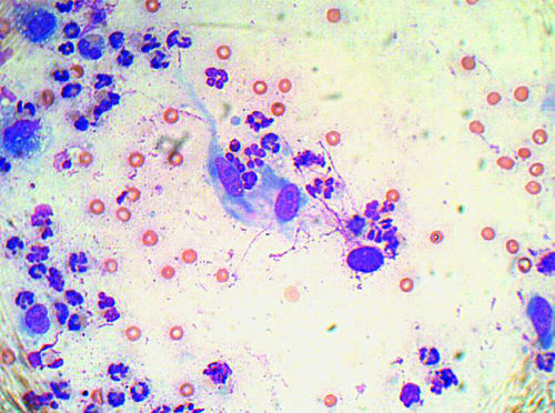
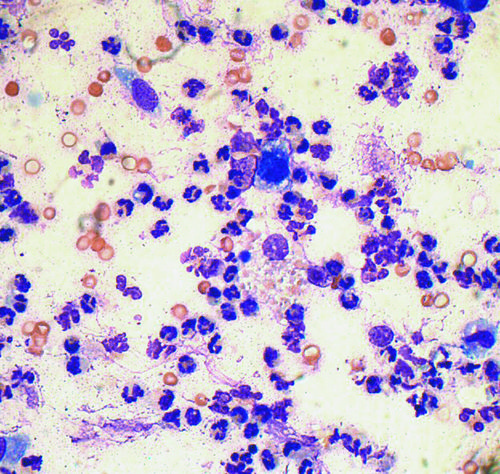
A seven-year-old neutered male Jack Russell Terrier presented with an interdigital mass of several weeks’ duration. Smears were made from an aspirate of the mass.
| Question | Answer | Article | |
| What features are illustrated in these photomicrographs (both Wright–Giemsa, ×50 oil)? |
|
Link to Article | |
| What is your interpretation of these findings? | The cytological features are consistent with moderate to marked pyogranulomatous inflammation with spindle cell proliferation, most likely fibroplasia. |
Link to Article | |
| What comments do you have regarding these findings? |
Atypical spindle cells with nucleoli and increased numbers typical of proliferation represent a diagnostic problem for all cytologists. |
Link to Article | |

