Difference between revisions of "Feline Medicine Q&A 16"
| (3 intermediate revisions by 3 users not shown) | |||
| Line 1: | Line 1: | ||
{{Template:Manson Sparkes}} | {{Template:Manson Sparkes}} | ||
| − | [[Image:|centre|500px]] | + | [[Image:Feline Medicine 16.jpg|centre|500px]] |
<br /> | <br /> | ||
| Line 12: | Line 12: | ||
|q1=What abnormalities are visible? | |q1=What abnormalities are visible? | ||
|a1= | |a1= | ||
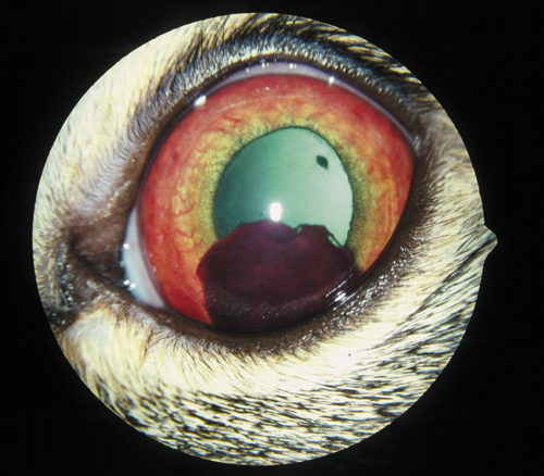
| − | There is obvious hyphaema (blood in the anterior chamber) which has clotted, suggesting a high fibrin content. The remaining view of the aqueous chamber is slightly hazy due to aqueous flare. The iris blood vessels are very prominent suggestive of an intense vasculitis. A focal deposit is visible on the anterior lens capsule and is likely to be a persistent pupillary membrane. The pupil has slightly irregular margins. | + | There is obvious hyphaema (blood in the anterior chamber) which has clotted, suggesting a high fibrin content. The remaining view of the aqueous chamber is slightly hazy due to aqueous flare. |
| − | |l1=Uveitis - Cat#Clinical Signs | + | |
| + | The iris blood vessels are very prominent suggestive of an intense vasculitis. A focal deposit is visible on the anterior lens capsule and is likely to be a persistent pupillary membrane. | ||
| + | |||
| + | The pupil has slightly irregular margins. | ||
| + | |l1=Uveitis - Cat and Dog#Clinical Signs | ||
|q2=What is the likely diagnosis? | |q2=What is the likely diagnosis? | ||
|a2= | |a2= | ||
Anterior uveitis (inflammation of the iris and/or ciliary body) with hyphaema. | Anterior uveitis (inflammation of the iris and/or ciliary body) with hyphaema. | ||
| − | |l2=Uveitis - Cat | + | |l2=Uveitis - Cat and Dog |
|q3=What are the possible causes of this condition? | |q3=What are the possible causes of this condition? | ||
|a3= | |a3= | ||
| Line 27: | Line 31: | ||
*Blunt and penetrating trauma to the eye. | *Blunt and penetrating trauma to the eye. | ||
FIP was diagnosed in this case. The cat had very high serum globulin levels (69 g/l [6.9 g/dl]), predominantly comprising gamma globulins on serum protein electrophoresis. Haematology revealed a mild anaemia, and lymphopenia. | FIP was diagnosed in this case. The cat had very high serum globulin levels (69 g/l [6.9 g/dl]), predominantly comprising gamma globulins on serum protein electrophoresis. Haematology revealed a mild anaemia, and lymphopenia. | ||
| − | |l3= | + | |l3=Feline Infectious Peritonitis |
</FlashCard> | </FlashCard> | ||
Latest revision as of 15:26, 10 October 2011
| This question was provided by Manson Publishing as part of the OVAL Project. See more Feline Medicine questions |
A 7-month-old DSH cat is presented with a 2-month history of waxing and waning inappetence, lethargy, and some weight loss. The owner has noticed that both of the eyes look abnormal.
| Question | Answer | Article | |
| What abnormalities are visible? | There is obvious hyphaema (blood in the anterior chamber) which has clotted, suggesting a high fibrin content. The remaining view of the aqueous chamber is slightly hazy due to aqueous flare. The iris blood vessels are very prominent suggestive of an intense vasculitis. A focal deposit is visible on the anterior lens capsule and is likely to be a persistent pupillary membrane. The pupil has slightly irregular margins. |
Link to Article | |
| What is the likely diagnosis? | Anterior uveitis (inflammation of the iris and/or ciliary body) with hyphaema. |
Link to Article | |
| What are the possible causes of this condition? | Differential diagnoses include:
FIP was diagnosed in this case. The cat had very high serum globulin levels (69 g/l [6.9 g/dl]), predominantly comprising gamma globulins on serum protein electrophoresis. Haematology revealed a mild anaemia, and lymphopenia. |
Link to Article | |
