Difference between revisions of "Small Animal Soft Tissue Surgery Q&A 04"
(No difference)
| |
Latest revision as of 12:10, 19 October 2011
| This question was provided by Manson Publishing as part of the OVAL Project. See more Small Animal Soft Tissue Surgery Q&A. |
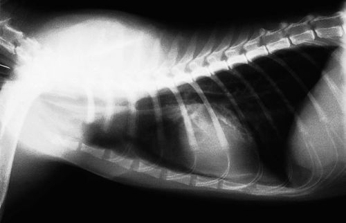
You are presented with a cat in severe respiratory distress. The cat was hit by a car two weeks previously and was treated by an emergency clinic. Because chest trauma was present, thoracic radiographs are made.
| Question | Answer | Article | |
| Based on the radiograph, what is your diagnosis? | Tracheal avulsion, rupture or transection are terms used to describe this condition. The intrathoracic trachea is transected. |
Link to Article | |
| Describe how this injury occurs. | The trachea is rapidly and severely stretched. The carina and lungs are fixed in their location and as the trachea is stretched, it tears just cranial to the carina. Tissues surrounding the trachea usually do not tear and pneumothorax does not occur. As healing progresses and scar tissue matures, stenosis or displacement of the trachea result in respiratory distress. |
Link to Article | |
| Does this cat most likely have inspiratory or expiratory dyspnea, and why? | Expiratory dyspnea is most prominent, because as the patient exhales, intrathoracic pressure increases and tends to collapse the pseudotrachea formed around the defect. During inspiration, there is negative intrathoracic pressure which tends to open this area. |
Link to Article | |
| Describe the surgical management of this problem. | The challenge for surgery is to maintain anesthesia and controlled ventilation during thoracotomy. The endotracheal tube must be long enough to reach the carina. It is wise to have a sterile endotracheal tube and breathing circuit which can be placed into the aborad portion of the trachea once the thoracotomy is performed. A right, 4th intercostal lateral thoracotomy is made. The azygous vein may need to be ligated for adequate exposure. The defect is identified and, if possible, the endotracheal tube placed per os is guided into the aborad segment. If the tube cannot be quickly advanced into the aborad segment, the pseudotracheal tissue is incised and a sterile endotracheal tube is inserted and connected to a sterile breathing circuit. The pseudotrachea is excised and the trachea prepared for anastomosis. The per os endotracheal tube is now advanced into the aborad segment and the trachea anastomosed in a routine manner. After anastomosis, if there is a leak, a mediastinal pleural patch is sutured around the anastomosis to provide adequate seal. |
Link to Article | |
