Difference between revisions of "Small Animal Orthopaedics Q&A 13"
Ggaitskell (talk | contribs) |
|||
| Line 1: | Line 1: | ||
| + | {{Manson | ||
| + | |book = Small Animal Orthopaedics Q&A}} | ||
| + | |||
[[File:SmAnOrth 13.jpg|centre|500px]] | [[File:SmAnOrth 13.jpg|centre|500px]] | ||
Revision as of 22:20, 23 October 2011
| This question was provided by Manson Publishing as part of the OVAL Project. See more Small Animal Orthopaedics Q&A. |
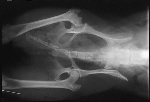
Ventrodorsal view radiograph of the pelvis of a ten-month-old, male West Highland White Terrier that has a nonweightbearing lameness of the left hindlimb.
| Question | Answer | Article | |
| Describe the radiographic abnormalities. | The normal trabecular pattern in the femoral neck region has been replaced by patches of increased radiodensity surrounded by areas of increased radiolucency. There is flattening of the articular surface of the femoral head, coxofemoral incongruity, and degenerative joint disease manifested by sclerosis of the subchondral bone in the region of the dorsal acetabular rim and increased radiodensity of the region of the acetabular fossa. These findings are consistent with avascular necrosis of the femoral head and secondary degenerative joint disease. |
[[|Link to Article]] | |
| Discuss the pathologic changes that occur with this condition. | The etiology of this condition has not been determined. Conjecture has centered on disruption of either the arterial or venous intraosseous and subsynovial blood flow of the femoral head and neck. Osteonecrosis occurs with ensuing trabecular resorption, fibroplasia and attempts at revascularisation. Trabecular weakness leads to subchondral bone collapse, formation of clefts and fissures in the articular cartilage, and structural deformation of the femoral head resulting in joint incongruity. Secondary generalized degenerative changes of the acetabulum also develop. |
[[|Link to Article]] | |
| What would be the most appropriate treatment for this dog? | Femoral head and neck excision is indicated for dogs with pain, lameness, and structural deformity of the femoral head and affords good limb function in approximately 75% of cases. Management of the dogs during the post-operative convalescent period is critical for a suc cessful outcome as recovery is often prolonged. Attention should be given to the use of post-operative analgesia and anti-inflammatory drugs. Physiotherapy, consisting of passive flexion and extension of the coxofemoral joint, slow walks on a leash and swimming, is also critical to achieving good limb function. |
[[|Link to Article]] | |
