Difference between revisions of "Equine Orthopaedics and Rheumatology Q&A 08"
| Line 17: | Line 17: | ||
The peroneus tertius is an almost completely tendinous structure that originates from the extensor fossa of the distal femur, runs over the cranial aspect of the tibia and inserts, after dividing into two, on the fibular, third and fourth tarsal bones and proximal third metatarsus. <br><br> | The peroneus tertius is an almost completely tendinous structure that originates from the extensor fossa of the distal femur, runs over the cranial aspect of the tibia and inserts, after dividing into two, on the fibular, third and fourth tarsal bones and proximal third metatarsus. <br><br> | ||
It is an important part of the reciprocal apparatus, which mechanically flexes the hock when the stifle joint is flexed. | It is an important part of the reciprocal apparatus, which mechanically flexes the hock when the stifle joint is flexed. | ||
| − | |l2= | + | |l2=Stay Apparatus - Horse Anatomy#The Stay Apparatus |
|q3=How does the injury occur? | |q3=How does the injury occur? | ||
|a3= The injury occurs as a result of extension of the hock as the stifle flexes. This can happen when an animal: <br> | |a3= The injury occurs as a result of extension of the hock as the stifle flexes. This can happen when an animal: <br> | ||
Latest revision as of 21:00, 31 October 2012
| This question was provided by Manson Publishing as part of the OVAL Project. See more Equine Orthopaedic and Rheumatological questions |
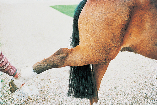
The figure illustrates a 13-year-old show jumping pony with a moderate, sudden onset lameness of the right hindlimb which developed after a fall in a competition.
| Question | Answer | Article | |
| What structure has been damaged? | The peroneus tertius has ruptured. The classical signs shown here are extension of the hock with the stifle flexed, and ‘puckering’ of the Achilles tendon (gastrocnemius and superficial digital flexor tendons).
|
Link to Article | |
| What is its function? | The peroneus tertius is an almost completely tendinous structure that originates from the extensor fossa of the distal femur, runs over the cranial aspect of the tibia and inserts, after dividing into two, on the fibular, third and fourth tarsal bones and proximal third metatarsus. |
Link to Article | |
| How does the injury occur? | The injury occurs as a result of extension of the hock as the stifle flexes. This can happen when an animal:
|
Link to Article | |
| How would you treat this pony, and what is its long-term prognosis? | There is no suitable surgical treatment. Complete box rest for 6–8 weeks, followed by a graduated exercise programme over the next 8–12 weeks, is the treatment of choice. The prognosis is fair to good. Healing is by scar tissue formation between the ruptured tendon ends. Return to a functional unit may not occur, or the peroneus tertius may be injured when the animal returns to work. |
Link to Article | |
