Difference between revisions of "Ornamental Fish Q&A 19"
m (Typo) |
|||
| (3 intermediate revisions by 3 users not shown) | |||
| Line 1: | Line 1: | ||
| + | {{Manson | ||
| + | |book = Ornamental Fish Q&A}} | ||
| + | |||
[[File:Ornamental Fish 19b.jpg|centre|500px]] | [[File:Ornamental Fish 19b.jpg|centre|500px]] | ||
| + | <br> | ||
| + | [[File:Ornamental Fish 19c.png|centre|500px]] | ||
<br> | <br> | ||
[[File:Ornamental Fish 19a.jpg|centre|500px]] | [[File:Ornamental Fish 19a.jpg|centre|500px]] | ||
| Line 23: | Line 28: | ||
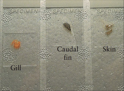
The second picture shows prepared gill, fin, and skin biopsies. | The second picture shows prepared gill, fin, and skin biopsies. | ||
| − | |l2= | + | |l2= Skin Scrape Procedure#Skin Scraping in Fish |
</FlashCard> | </FlashCard> | ||
Latest revision as of 15:34, 17 July 2018
| This question was provided by Manson Publishing as part of the OVAL Project. See more Ornamental Fish Q&A. |
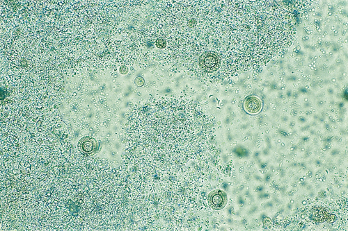
These rapidly spinning ciliated organisms were seen on the examination of a skin scrape (×100).
| Question | Answer | Article | |
| Name the organism. | Trichodina, a ciliated protozoan parasite which is a common cause of skin and gill disease in ornamental fish. |
Link to Article | |
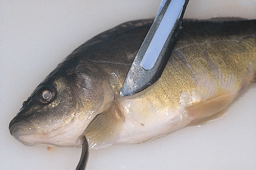
| How is a skin scraping prepared? | A skin scrape is prepared by obtaining a small quantity of mucus from the skin surface of a freshly dead or anesthetized fish. Areas of predilection for skin parasites are those adjacent to the fins and it is a good approach to obtain material from these sites. The mucus is suspended in a small quantity of water on a microscope slide (preferably pond or tank water from which the fish was taken; tap or distilled water may destroy the parasites), a cover slip applied, and the preparation examined under the microscope (×40 magnification should be sufficient to visualize most parasites). The second picture shows prepared gill, fin, and skin biopsies. |
Link to Article | |


