Difference between revisions of "Small Animal Emergency and Critical Care Medicine: Self-Assessment Color Review, Second Edition, Q&A 21"
Jump to navigation
Jump to search
(Created page with "{{CRC Press}} {{Student tip |X = showing what is normal/abnormal, and when intervention is required. }} <br><br><br> File:Small Animal Emergency and Critical Care Medicine 2...") |
|||
| Line 28: | Line 28: | ||
[[Category:CRC Press flashcards]] | [[Category:CRC Press flashcards]] | ||
| − | To purchase the full text with your 20% discount, go to the [https://www.crcpress.com/9781482225921 CRC Press] Veterinary website | + | To purchase the full text with your 20% off discount code, go to the [https://www.crcpress.com/9781482225921 CRC Press] Veterinary website. |
| − | |||
Revision as of 18:04, 6 November 2018
| This question was provided by CRC Press. See more case-based flashcards |

|
Student tip: This case is showing what is normal/abnormal, and when intervention is required. |
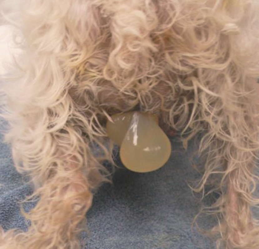
A 4-year-old female entire Schnauzer-cross presents because of two fluid-filled sacs hanging from her vulva (202). The first sac was observed about 45 minutes prior to presentation and the second sac 20 minutes prior to presentation. She was bred approximately 62 days ago to a large male Schnauzer. This is her first litter. T = 37.7°C (99.9°F); HR = 150 bpm; RR = 60 bpm; CRT = 1 sec; MM pale pink, moist. Abdomen is tense and she is having visible abdominal con-tractions, which were not observed prior to presentation. Fetal HR = 180 bpm. Rectal examination reveals a fetus at the pelvic inlet.
| Question | Answer | Article | |
| What are the fetal and maternal causes of dystocia? | Fetal causes include increased fetal size, abnormal presentation, position, posture, or development. Maternal causes include systemic disease, reduced pelvic size, abnormal reproductive tract anatomy, or abnormal expulsion (uterine intertia).
|
Link to Article | |
| List at least four signs of dystocia. | Presence of uteroverdin without immediate fetal delivery; prepartum pregnancy toxemia (ketosis without hyperglycemia); failure to produce a fetus after 30 minutes of strong contractions; weak straining for 2 hours without delivery; >4–6 hours since birth of last pup; retained pup in canal.
|
Link to Article | |
| Stage 1 labour is defined by a progesterone level less than what value? | <2 ng/ml.
|
Link to Article | |
| What is oxytocin, what is its mechanism of action, and what are the pros and cons of its use in a pet with dystocia? | A drug that promotes uterine contraction (ecbolic) by increasing intracellular influx of calcium. It can be used for non-obstructive dystocia, when uterine inertia is not complete. Pros: stimulates uterine contraction, reduces uterine hemorrhage, promotes uterine involution, and decreases incidence of fetal membrane retention. Cons: can also interrupt uteroplacental blood flow and cause ineffective tetanic uterine contractions, which could affect fetal viability. Higher doses of oxytocin are associated with fetal hypoxemia; conservative doses (0.25–2 IU/dog SC) are recommended. Oxytocin is avoided when an immediate cesarean section can be performed to improve fetal viability.
|
Link to Article | |
| List six indications for emergency cesarean section. | 1) Obstruction: fetal–maternal disparity, fetal malposition, non-fetal obstruction of canal, uterine torsion. (2) Fetal distress (HR <160 bpm). (3) Failure to deliver with oxytocin (non-obstructive dystocia). (4) Presence of uteroverdin, meconium, excessive hemorrhage, or pus in vaginal discharge. (5) Maternal illness. (6) >65 days post breeding (or 63 days post ovulation).
|
Link to Article | |
To purchase the full text with your 20% off discount code, go to the CRC Press Veterinary website.
