Difference between revisions of "Small Animal Emergency and Critical Care Medicine: Self-Assessment Color Review, Second Edition, Q&A 19"
Jump to navigation
Jump to search
| Line 28: | Line 28: | ||
[[Category:CRC Press flashcards]] | [[Category:CRC Press flashcards]] | ||
| − | To purchase the full text with your 20% discount, go to the [https://www.crcpress.com/9781482225921 CRC Press] Veterinary website | + | To purchase the full text with your 20% off discount code, go to the [https://www.crcpress.com/9781482225921 CRC Press] Veterinary website. |
| − | |||
Revision as of 16:15, 7 November 2018
| This question was provided by CRC Press. See more case-based flashcards |

|
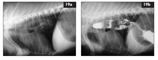
Student tip: This case is showing radiographs demonstrating plain vs barium study. |
A 6-year-old female spayed Maltese presents for lethargy and vomiting or regurgitating a small amount of water with a piece of rawhide in it. She started howling in the car. T = 36.9°C (98.4°F); HR = 80 bpm; RR = 45 bpm; CRT = 2 sec; MM pink, slightly dry; pulses normal; perfusion normal; 5% dehydrated. Abdominal and thoracic examination normal. The dog is hypersalivating. Plain (19a) and barium contrast (19b) lateral thoracic radiographs are obtained.
| Question | Answer | Article | |
| Interpret the radiographs and provide a radiographic diagnosis. | There is a soft tissue density between the thoracic inlet and the cranial aspect of the heart on the plain radiograph. The barium contrast highlights dilation of the esophagus along with a bi-lobed filling defect dorsal to the heart, a portion passing through the stomach and into the upper duodenum. There is gas dilation of the stomach. This is consistent with an esophageal FB causing a partial obstruction and aerophagia.
|
Link to Article | |
| What are the pros and cons of barium administration? What are the alternative imaging options? | Pros: barium can coat ulcers in the stomach, has anti-secretory effects, and may promote GI motility. Cons: leakage of barium into the mediastinum, trachea, or pleural cavity if esophageal perforation is present; aspiration of barium into the lungs; poor visualization if endoscopy performed after barium administration. Alternative imaging: flexible endoscopic or rigid orogastic tube evaluation; CT.
|
Link to Article | |
| What are the common locations for the problem identified? | Cranial to the thoracic inlet, cranial to the heart, and cranial to the diaphragm (esophageal hiatus).
|
Link to Article | |
| What are some consequences of this problem? | Esophageal erosion and ulceration are common; esophageal perforation possibly leading to pneumomediastinum or (tension) pneumothorax and pyothorax; esophageal stricture may occur long term.
|
Link to Article | |
| What are your treatment goals? How would you correct this problem? | To stabilize the patient and remove the FB obstruction while preserving the integrity of the GI tract. Treatment for the consequences of the lodged FB is planned after removal of the obstruction and examination of the esophagus. There are several methods for removal: (1) endoscopic retrograde removal (if possible); (2) endoscopic or orogastric tube-assisted normograde advancement of the FB into the stomach for digestion (if possible) or gastrotomy; or (3) surgical intervention – some caudal esophageal FBs can be surgically removed through an abdominal approach with gastrotomy; others (like this one) require a thoracotomy.
|
Link to Article | |
To purchase the full text with your 20% off discount code, go to the CRC Press Veterinary website.
