Difference between revisions of "Rabbit Medicine and Surgery: Self-Assessment Color Review, Second Edition, Q&A 01"
(Created page with "{{CRC Press}} {{Student tip |X = an example with great pictures, with clear explanation of each one in the answers }} <br><br><br> File: Rabbit Medicine and Surgery Q1.PNG|...") |
|||
| Line 17: | Line 17: | ||
</FlashCard> | </FlashCard> | ||
| − | To purchase the full text with your 20% off discount code, go to the [https://www.crcpress.com/9781498730792 CRC Press] Veterinary website | + | To purchase the full text with your 20% off discount code, go to the [https://www.crcpress.com/9781498730792 CRC Press] Veterinary website. |
[[Category:CRC Press flashcards]] | [[Category:CRC Press flashcards]] | ||
Revision as of 10:51, 22 November 2018
| This question was provided by CRC Press. See more case-based flashcards |

|
Student tip: This case is an example with great pictures, with clear explanation of each one in the answers |
Examine the images above.
| Question | Answer | Article | |
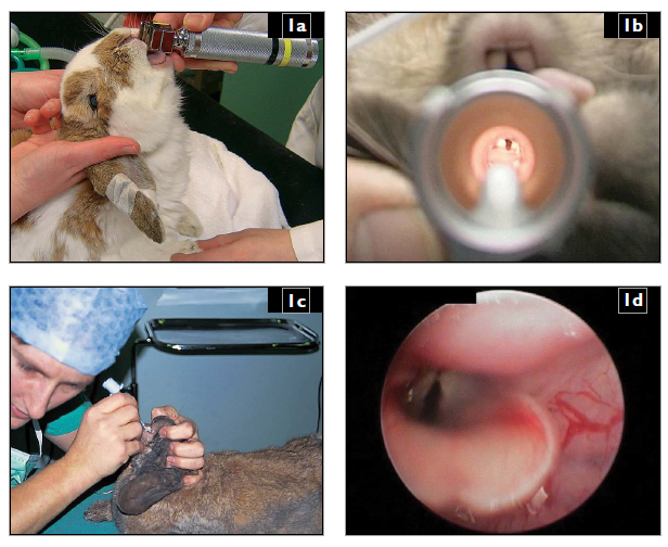
| Describe the techniques available for endotracheal intubation of an anaesthetized rabbit (1a, b, c, d)? | 1. DIRECT VISUALIZATION. Place the anaesthetized rabbit in sternal recumbency, extend the neck vertically by elevating the head, grasp the tongue gently and retract it through the diastema and hold to one side. Visualize the larynx using a Wisconsin size 1 laryngoscope blade (1a) and insert a 2.5–3.0 mm endotracheal tube (1b). Alternatively, position the rabbit as above, visualize the larynx using an otoscope or Wisconsin size 1 laryngoscope blade or similar, place an introducer (e.g. 3–5 Fr urinary catheter) into the larynx through an otoscope or over a laryngoscope, remove the otoscope/laryngoscope and introduce an endotracheal tube gently over the introducer and then remove the introducer. A further alternative is to use the otoscope and place the endotracheal tube with its connector removed directly through the otoscope cone, replacing the connector after removal of the otoscope. The larynx can also be visualized using a small rigid endoscope while the endotracheal tube is passed alongside it into the larynx. All the above methods can also be performed with the rabbit in dorsal recumbency with the neck extended.
|
Link to Article | |
To purchase the full text with your 20% off discount code, go to the CRC Press Veterinary website.
