Difference between revisions of "Rabbit Medicine and Surgery: Self-Assessment Color Review, Second Edition, Q&A 08"
Jump to navigation
Jump to search
(Created page with "{{CRC Press}} <br><br><br> centre <br> ''A lateral abdominal radiograph from a rabbit is shown above.'' <br> <br> <FlashCard que...") |
|||
| Line 12: | Line 12: | ||
</FlashCard> | </FlashCard> | ||
| − | To purchase the full text with your 20% off discount code, go to the [https://www.crcpress.com/9781498730792 CRC Press] Veterinary website | + | To purchase the full text with your 20% off discount code, go to the [https://www.crcpress.com/9781498730792 CRC Press] Veterinary website. |
[[Category:CRC Press flashcards]] | [[Category:CRC Press flashcards]] | ||
Revision as of 11:08, 22 November 2018
| This question was provided by CRC Press. See more case-based flashcards |
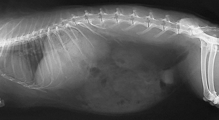
A lateral abdominal radiograph from a rabbit is shown above.
| Question | Answer | Article | |
| What normal anatomical features can be seen? | Normal abdominal contents that may be easily visualized include the simple stomach, intestines (duodenum, long caecum with appendix, large intestine), kidneys, liver and bladder. Food and caecal pellets are always present in the stomach. The caecum lies on the right side of the abdomen. Rabbits often have large amounts of retroperitoneal fat, which displaces the kidney ventrally. In intact female rabbits the uterus may be visible situated ventral to the large intestine and dorsal to the bladder. The bladder may normally contain a small number of calcium carbonate crystals, which will appear radiodense, as is evident in this radiograph.
|
Link to Article | |
To purchase the full text with your 20% off discount code, go to the CRC Press Veterinary website.
