Difference between revisions of "Canine Infectious Diseases: Self-Assessment Color Review, Q&A 09"
(No difference)
| |
Latest revision as of 09:44, 26 November 2018
| This question was provided by CRC Press. See more case-based flashcards |

|
Student tip: This case is an example using a good image and helpful use of blood results |
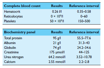
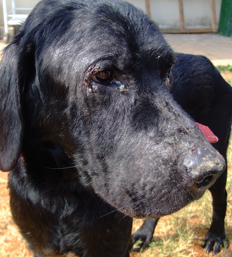
A 6-year-old male Labrador Retriever from Bristol, UK, that had lived in Spain in recent years before returning to the UK 6 months ago was evaluated for a skin problem that included alopecia, crusts, scaling, and hyperpigmentation around the face, ears, limbs, and dorsum (see image). Over the last 2 months, the dog had developed progressive inappetence, lethargy, polydipsia, and weight loss. The dog had regularly received core vaccines (distemper virus, adenovirus, parvovirus, rabies virus, and Leptospira spp.) and was treated regularly for intestinal parasites. Physical examination revealed a thin body condition (BCS 2–3/9), mucoid ocular discharge, generalized peripheral lymphadenomegaly, splenomegaly, mild pyrexia, and pale mucous membranes. A CBC, biochemistry panel, and urinalysis were performed. Urinalysis revealed a specific gravity of 1.015, with 3+ protein and a benign sediment. The UPCR was 1.5 (RI <1.5).
| Question | Answer | Article | |
| What are the most likely differential diagnoses in this dog? | n this case, exfoliative cutaneous lesions, alopecia, and systemic signs were observed. There was also a mild anemia, hyperproteinemia, hyperglobulinemia, and hypoalbuminemia, suggesting chronic immune stimulation. Renal failure was evidenced by the azotemia combined with isosthenuria. The increased UPCR together with a benign urine sediment raised concern for the possibility of glomerular disease. Differential diagnoses include chronic inflammatory diseases with dermatologic involvement, such as leishmaniosis, immune-mediated diseases (especially systemic lupus erythematosus), and neoplasia (e.g. cutaneous lymphoma).
|
Link to Article | |
| What diagnostic tests should be done next? | Further diagnostic tests to confirm a diagnosis of leishmaniosis include cytologic examination of impression smears of the skin lesions; biopsy of the skin lesions; fine needle aspiration and cytology of the lymph nodes; antibody tests for Leishmania spp.; or PCR of skin, blood, conjunctival swabs, or bone marrow. To diagnose immune-mediated diseases, antinuclear antibody tests or skin biopsies might be indicated. Skin biopsies would also be required for diagnosis of cutaneous lymphoma.
|
Link to Article | |
To purchase the full text with your 20% discount, go to the CRC Press Veterinary website and use code VET18.

