Difference between revisions of "Canine Infectious Diseases: Self-Assessment Color Review, Q&A 13"
Jump to navigation
Jump to search
(No difference)
| |
Latest revision as of 09:45, 26 November 2018
| This question was provided by CRC Press. See more case-based flashcards |

|
Student tip: This case is a demonstration of excellent echocardiography imagery |
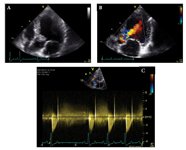
Case 13 is the same dog as in Case 12. Echocardiography was performed. A left apical five-chamber view (A), a colour Doppler image (B), and a CW Doppler image (C) are shown.
| Question | Answer | Article | |
| What is your interpretation of the echocardiographic images? | Figure A shows a hyperechogenic vegetative oscillating lesion on the septal aortic valve cusps. The left ventricle appears slightly volume overloaded. Figure B shows a severe aortic regurgitation jet in diastole, approaching the center of the left ventricle, due to a vegetative lesion on the septal cusps. This explains the diastolic murmur. Figure C shows the continuous wave Doppler of the aortic velocity. The systolic velocity is about 4 m/s, which resembles a pressure gradient of 64 mmHg according to the modified Bernoulli equation (4 × v2). Normal velocity is <2.0 m/s, so there is evidence of moderate aortic stenosis. This explains the systolic component of the murmur.
|
Link to Article | |
| What is your main differential diagnosis? | Taking the echocardiographic findings together, there is a high suspicion of aortic valve endocarditis causing a systolic and diastolic murmur due to aortic stenosis and aortic valve insufficiency. Both murmurs together auscultate as a continuous murmur. The main underlying reason could be congenital aortic stenosis. In this dog, the aortic valve is severely thickened and there is a vegetative lesion, which makes congenital aortic stenosis less likely. However, aortic valve stenosis can be a predisposing factor for the development of endocarditis. Although many dogs with endocarditis are presented with a history of fever (60%), this dog did not have fever at the time of evaluation.
|
Link to Article | |
| What further tests would you recommend? | Other diagnostic tests that could be performed in this dog to assess the severity of disease and determine whether there are complications of endocarditis (such as thromboembolic disorders) include evaluation of laboratory parameters (CBC, biochemistry panel, and urinalysis), thoracic radiographs, abdominal ultrasound, urinalysis, and bacterial urine culture, bacterial blood cultures (three specimens from different sites), and diagnostic tests for Bartonella spp.
|
[[ Replace text with name and subsection of relevant WikiVet page if in existence eg. Feather - Anatomy & Physiology#Structure & Function |Link to Article]] | |
To purchase the full text with your 20% discount, go to the CRC Press Veterinary website and use code VET18.
