Difference between revisions of "Canine Infectious Diseases: Self-Assessment Color Review, Q&A 18"
Jump to navigation
Jump to search
(No difference)
| |
Latest revision as of 09:46, 26 November 2018
| This question was provided by CRC Press. See more case-based flashcards |

|
Student tip: This case is a good explanation, developing the previous Case (17) |
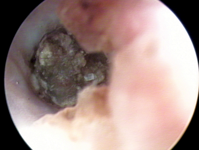
Case 18 is the same dog as case 17. Otoscopic evaluation was performed under sedation and revealed exudate in the left horizontal ear canal (see image). The tympanic membranes could not be examined because of severe ear canal stenosis. Open-mouth anterior–posterior radiographs of the skull revealed a bilateral thickening of the bullae that was more severe on the left side.
| Question | Answer | Article | |
| What is your diagnosis? | The dog has chronic bilateral otitis externa and otitis media. The left ear is more severely affected and there is likely to be secondary otitis media, based on the clinical signs present.
|
Link to Article | |
| What additional diagnostic tests might be useful to better understand the condition? | Otitis media is frequently caused by extension of otitis externa. Otitis externa is the consequence of a primary skin disease promoting bacterial and yeast overgrowth. The most common underlying disease in dogs is allergic dermatitis, but foreign bodies, hypothyroidism, mite infestations, and cornification disorders (such as seborrhea in Cocker Spaniels) can also predispose to otitis externa. Bacterial infections (such as those caused by Staphylococcus pseudintermedius, Pseudomonas aeruginosa) and yeast (Malassezia) infections are perpetuating factors, and the chronic inflammation produces progressive pathologic changes in the external ear, including the tympanic membrane. Therefore, the underlying cause of chronic or recurrent otitis externa should be investigated and treated if possible. Cytology and culture (ideally of material obtained from the middle ear) are useful to guide the choice of systemic antimicrobial drug therapy. Cytology of both external ear canals should be performed, at a minimum, to confirm the presence of bacteria and yeast together with neutrophils.
|
Link to Article | |
| What treatment is indicated? | Ear flushing under anesthesia should be recommended. A 3–4-day course of systemic glucocorticoids can reduce stenosis due to inflammation and make otoscopic evaluation without anesthesia, ear cleaning, and topical treatment possible. Owners should be educated regarding the complications of ear canal flushing (e.g. Horner’s syndrome, facial nerve paralysis, vestibular signs, and deafness). Topical combination therapy with glucocorticoids, antibiotics, and antifungal drugs is then indicated to control infection, pain, pruritus, and inflammation. A variety of combination preparations are available and selection depends largely on clinician preference. In case of Pseudomonas aeruginosa infection, topical treatment with fluoroquinolones can be potentiated by combination therapy with topical Tris-EDTA or dexamethasone solutions. In severe cases, systemic glucocorticoids and antimicrobial drugs can be combined with topical therapy. When there are advanced pathologic changes of the ear canal (stenosis and calcification) and otitis media, total ear canal ablation and bulla osteotomy might be required. Addressing the underlying causes is also recommended when possible.
|
Link to Article | |
To purchase the full text with your 20% discount, go to the CRC Press Veterinary website and use code VET18.
