Difference between revisions of "Hyoid Apparatus - Anatomy & Physiology"
Fiorecastro (talk | contribs) |
Fiorecastro (talk | contribs) |
(No difference)
| |
Latest revision as of 17:31, 7 November 2022
Introduction
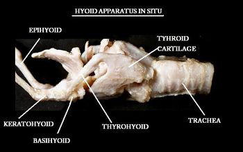
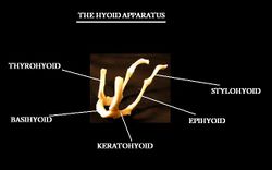
The hyoid apparatus holds the larynx in place and supports the pharynx and tongue from the skull. It is made up of 5 different bones, which vary in length and size depending on the species.
Structure and Function
It is attached to the temporal region of the skull by a synchondrosis joint. It is palpable through the pharynx and is visible when the pharynx is viewed through the mouth. The basihyoid is palpable within the intermandibular space.
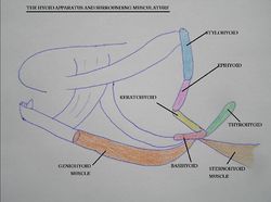
The sternohyoid muscle pulls the hyoid caudally and the geniohyoid muscle pulls the hyoid rostrally.
The hyoid bones
1. Basihyoid - Unpaired bone.
2. Stylohyoid - Articulates with base of skull at the petrus temporal. A paired bone.
3. Epihyoid - A paired bone.
4. Keratohyoid - A paired bone.
5. Thyrohyoid - Articulates with the thyroid cartilage of the larynx. A paired bone.
Species Differences
Carnivores
Stylohyoid bones are not palpable.
Equine
Epihyoid is small, the lingual process is present and they have a well developed stylohyoid muscle.
Ruminants
Lingual process is present.
Porcine
Lingual process is present.
| Hyoid Apparatus - Anatomy & Physiology Learning Resources | |
|---|---|
 Test your knowledge using flashcard type questions |
Hyoid apparatus anatomy |
 Selection of relevant videos |
Ventral muscles of the head potcast Hyoid apparatus potcast |
Webinars
Failed to load RSS feed from https://www.thewebinarvet.com/gastroenterology-and-nutrition/webinars/feed: Error parsing XML for RSS


