Difference between revisions of "Hoof - Horse Anatomy"
Fiorecastro (talk | contribs) |
|||
| (8 intermediate revisions by 3 users not shown) | |||
| Line 1: | Line 1: | ||
| − | {{ | + | {{OpenPagesTop}} |
==Introduction== | ==Introduction== | ||
The general anatomy of the hoof can be found [[Hoof - Anatomy & Physiology|here]]. The following section will focus on the equine hoof specifics. | The general anatomy of the hoof can be found [[Hoof - Anatomy & Physiology|here]]. The following section will focus on the equine hoof specifics. | ||
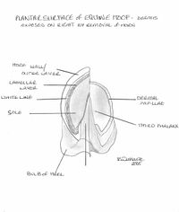
| + | [[image: Plantar hoof aspect.jpg|thumb|200px|right|A view of the solar surface of an equine hoof. The wall has been removed on the right to show the underlying dermis. ©Rachael Wallace2008]] | ||
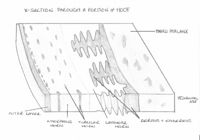
| + | [[image: X-section through hoof.jpg|thumb|200px|right|A X-section through a typical hoof. ©Rachael Wallace2008]] | ||
| + | The hoof encloses the corium (dermis), digital cushion, [[Phalanges - Horse Anatomy#Distal Phalanx|distal phalanx]] , [[Joints and Ligaments - Horse Anatomy#Distal Interphalangeal (Coffin) Joint|distal interphalangeal]] (coffin) joint, the distal aspect of the [[Phalanges - Horse Anatomy#Middle Phalanx|middle phalanx]] , [[Phalanges - Horse Anatomy#Distal Sesamoid (Navicular) Bone|distal sesamoid]] (navicular) bone, ligaments and the insertion points of the [[Tendons - Horse Anatomy#Extensors|common digital extensor tendon]] and [[Tendons - Horse Anatomy#Flexors|deep digital flexor tendon]]. | ||
| − | + | The hooves in newborn foals are bilaterally symmetrical. Over a period of just a few months, forces exerted on the hoof during locomotion cause a visible difference between the right and left, as well as front and hind hooves. Thus, isolated specimens of equine feet can be distinguished as follows: | |
| − | |||
| − | The hooves in newborn foals are bilaterally symmetrical. Over a period of just a few months, forces exerted on the hoof during locomotion cause a visible difference between the right and left, as well as front and hind hooves. Thus, isolated specimens of | ||
* Front vs hind: | * Front vs hind: | ||
**''Front'': The angle between the '''toe''' and the ground is approximately 45 degrees. The sole is circular in shape. | **''Front'': The angle between the '''toe''' and the ground is approximately 45 degrees. The sole is circular in shape. | ||
| Line 13: | Line 14: | ||
==Topographical Anatomy== | ==Topographical Anatomy== | ||
| − | The equine hoof can be divided into three topographical regions; the '''wall''', the '''frog''' and the '''sole'''. | + | The equine hoof can be divided into three topographical regions; the '''wall''', the '''frog''' and the '''sole'''. A well-trimmed foot should weight bear on its walls, bars and frog. This occurs as the weight applied to the [[Phalanges - Horse Anatomy#Distal Phalanx|distal phalanx]] is then transferred across the interdigitating laminae to the hoof wall. Thus an injury resulting in damage to the laminae is of extreme importance to the horse. |
| + | |||
===Wall=== | ===Wall=== | ||
The '''wall''' forms the medial, lateral and dorsal aspect of the hoof and it can be further divided into the '''toe''', '''quarters''' and '''heels'''. At the heel the walls reflect back on themselves at a point called the '''angles''' and in doing so forms the '''bars'''. The bars, although moving cranially, gradually fade along the edge of the frog and never actually meet. | The '''wall''' forms the medial, lateral and dorsal aspect of the hoof and it can be further divided into the '''toe''', '''quarters''' and '''heels'''. At the heel the walls reflect back on themselves at a point called the '''angles''' and in doing so forms the '''bars'''. The bars, although moving cranially, gradually fade along the edge of the frog and never actually meet. | ||
| + | |||
===Frog=== | ===Frog=== | ||
The '''frog''' is a wedge-shaped structure which sits between the bars and has an apex facing distally, with 2 crura flanking a central sulcus. Between the crus and bar of each half of the sole lies the '''collateral sulcus'''. Opposite the apex, the frog expands forming the '''bulbs of the heel'''. The frog is a mass of keratinized stratified squamous epithelium, which is softer than other parts of the hoof due to its increased water content. Usually, the frog contributes to the weightbearing surface where it functions as a shock absorber. Apocrine glands within the corium of the frog produce secretions on the surface. | The '''frog''' is a wedge-shaped structure which sits between the bars and has an apex facing distally, with 2 crura flanking a central sulcus. Between the crus and bar of each half of the sole lies the '''collateral sulcus'''. Opposite the apex, the frog expands forming the '''bulbs of the heel'''. The frog is a mass of keratinized stratified squamous epithelium, which is softer than other parts of the hoof due to its increased water content. Usually, the frog contributes to the weightbearing surface where it functions as a shock absorber. Apocrine glands within the corium of the frog produce secretions on the surface. | ||
| Line 21: | Line 24: | ||
===Sole=== | ===Sole=== | ||
The '''sole''' is the area distal to the bars and apex of the frog enclosed by the hoof wall. The area where the bars and wall enclose it is known as the '''angle of the sole'''. Since the sole is slightly concave, the majority of the horse's weight is transferred through the margin of the sole. | The '''sole''' is the area distal to the bars and apex of the frog enclosed by the hoof wall. The area where the bars and wall enclose it is known as the '''angle of the sole'''. Since the sole is slightly concave, the majority of the horse's weight is transferred through the margin of the sole. | ||
| − | |||
| − | |||
| − | |||
== Deeper Structures of the Hoof== | == Deeper Structures of the Hoof== | ||
| − | The dermis of the distal phalanx is arranged in hundreds of leaves or '''laminae''', each of which has microscopic '''secondary laminae'''. The coronary region has a germinative layer associated with papillae that is responsible for producing the horn tubules that make up the hoof wall. This wall glides distally at a rate of 5-6mm a month and by forming epidermal laminae itself it interdigitates with the underlying dermal laminae. Neither of these laminae are pigmented so when the epidermal laminae appear on the solar surface, a non-pigmented region known as the '''white line''' appears. The white line is used as important landmark in farriery as structures central to the line will be dermal and so vascular and sensitive. | + | The dermis of the [[Phalanges - Horse Anatomy#Distal Phalanx|distal phalanx]] is arranged in hundreds of leaves or '''laminae''', each of which has microscopic '''secondary laminae'''. The coronary region has a germinative layer associated with papillae that is responsible for producing the horn tubules that make up the hoof wall. This wall glides distally at a rate of 5-6mm a month and by forming epidermal laminae itself it interdigitates with the underlying dermal laminae. Neither of these laminae are pigmented so when the epidermal laminae appear on the solar surface, a non-pigmented region known as the '''white line''' appears. The white line is used as important landmark in farriery as structures central to the line will be dermal and so vascular and sensitive. |
The dermis in the frog is also arranged in papillae and produces incompletely keratinised flexuous horn tubules resulting in a soft, elastic horn. The sole area also has papillae that produces superficially flakey horn. The coronary part of the wall is surrounded by a bony prominence called the '''periople'''. This soft, lightly coloured area is restricted to this proximal area and is produced by the germative layer covering the papillae. The rest of the hoof is covered by the '''tectorial layer''', this is a very thin layer of horn that is covered distally by the growth of the horn. | The dermis in the frog is also arranged in papillae and produces incompletely keratinised flexuous horn tubules resulting in a soft, elastic horn. The sole area also has papillae that produces superficially flakey horn. The coronary part of the wall is surrounded by a bony prominence called the '''periople'''. This soft, lightly coloured area is restricted to this proximal area and is produced by the germative layer covering the papillae. The rest of the hoof is covered by the '''tectorial layer''', this is a very thin layer of horn that is covered distally by the growth of the horn. | ||
===Digital Cushion=== | ===Digital Cushion=== | ||
| − | The digital cushion lies between the ungual cartilages and is collagenous, elastic tissue infiltrated by adipose tissue. At the bulbs of the heel, it is subcutaneous and is soft and loose in texture. The flexible material that forms the digital cushion is produced from the hypodermis of the frog. It acts as one of the major shock absorbers of the foot. It is connected dorsoproximally with the digital annular ligament and attached at the apex to the deep digital flexor tendon at its point of insertion on the distal phalanx. | + | The digital cushion lies between the ungual cartilages and is collagenous, elastic tissue infiltrated by adipose tissue. At the bulbs of the heel, it is subcutaneous and is soft and loose in texture. The flexible material that forms the digital cushion is produced from the hypodermis of the frog. It acts as one of the major shock absorbers of the foot. It is connected dorsoproximally with the digital annular ligament and attached at the apex to the [[Tendons - Horse Anatomy#Flexors|deep digital flexor tendon]] at its point of insertion on the [[Phalanges - Horse Anatomy#Distal Phalanx|distal phalanx]]. |
===Ungual Cartilages=== | ===Ungual Cartilages=== | ||
| − | The medial and lateral cartilages of the distal phalanx (ungual cartilages) extend from the palmar processes of the bone to the coronary region of the hoof. They can be palpated proximally. The axial surface is concave and their abaxial surface convex. They curve towards each other in the heel region and become thicker distally. The cartilages contain several foraminae through which veins pass from the palmar venous plexus to the coronary venous plexus. | + | The medial and lateral cartilages of the [[Phalanges - Horse Anatomy#Distal Phalanx|distal phalanx]] (ungual cartilages) extend from the palmar processes of the bone to the coronary region of the hoof. They can be palpated proximally. The axial surface is concave and their abaxial surface convex. They curve towards each other in the heel region and become thicker distally. The cartilages contain several foraminae through which veins pass from the palmar venous plexus to the coronary venous plexus. |
| − | The ungual cartilages have a series of five ligaments going to the medial/lateral surfaces of the three phalanges and distal sesamoid (navicular) bone. A short ligament extends from the dorsal aspect to the dorsal surface of the middle phalanx. A thin elastic fibre extends from the proximal aspect to the proximal phalanx. Several short fibres attach from the distal part of the cartilages to the distal phalanx. Another ligament extends from the dorsal region of the cartilages to the insertion point of the deep digital flexor tendon. Finally, an extension of the collateral sesamoidean ligament attaches them to the navicular bone. | + | The ungual cartilages have a series of five ligaments going to the medial/lateral surfaces of the three phalanges and [[Phalanges - Horse Anatomy#Distal Sesamoid (Navicular) Bone|distal sesamoid]](navicular) bone. A short ligament extends from the dorsal aspect to the dorsal surface of the [[Phalanges - Horse Anatomy#Middle Phalanx|middle phalanx]]. A thin elastic fibre extends from the proximal aspect to the [[Phalanges - Horse Anatomy#Proximal Phalanx|proximal phalanx]]. Several short fibres attach from the distal part of the cartilages to the [[Phalanges - Horse Anatomy#Distal Phalanx|distal phalanx]]. Another ligament extends from the dorsal region of the cartilages to the insertion point of the [[Tendons - Horse Anatomy#Flexors|deep digital flexor tendon]]. Finally, an extension of the collateral sesamoidean ligament attaches them to the [[Phalanges - Horse Anatomy#Distal Sesamoid (Navicular) Bone|navicular bone]]. |
| + | {{Template:Learning | ||
| + | |OVAM = [http://oa.scopty.com 3D model of the horse's distal limb] | ||
| + | }} | ||
| + | ==Webinars== | ||
| + | <rss max="10" highlight="none">https://www.thewebinarvet.com/orthopaedics/webinars/feed</rss> | ||
| − | [[ | + | [[Category:To Do - AP Review]] |
| + | [[Category:Horse Anatomy]] | ||
Latest revision as of 16:56, 3 January 2023
Introduction
The general anatomy of the hoof can be found here. The following section will focus on the equine hoof specifics.
The hoof encloses the corium (dermis), digital cushion, distal phalanx , distal interphalangeal (coffin) joint, the distal aspect of the middle phalanx , distal sesamoid (navicular) bone, ligaments and the insertion points of the common digital extensor tendon and deep digital flexor tendon.
The hooves in newborn foals are bilaterally symmetrical. Over a period of just a few months, forces exerted on the hoof during locomotion cause a visible difference between the right and left, as well as front and hind hooves. Thus, isolated specimens of equine feet can be distinguished as follows:
- Front vs hind:
- Front: The angle between the toe and the ground is approximately 45 degrees. The sole is circular in shape.
- Hind: The angle between the toe and the ground is 50-55 degrees. The sole is oval in shape.
- Right vs left:
- Quarters (lateral and medial walls) are steeper on the medial side of the hoof.
Topographical Anatomy
The equine hoof can be divided into three topographical regions; the wall, the frog and the sole. A well-trimmed foot should weight bear on its walls, bars and frog. This occurs as the weight applied to the distal phalanx is then transferred across the interdigitating laminae to the hoof wall. Thus an injury resulting in damage to the laminae is of extreme importance to the horse.
Wall
The wall forms the medial, lateral and dorsal aspect of the hoof and it can be further divided into the toe, quarters and heels. At the heel the walls reflect back on themselves at a point called the angles and in doing so forms the bars. The bars, although moving cranially, gradually fade along the edge of the frog and never actually meet.
Frog
The frog is a wedge-shaped structure which sits between the bars and has an apex facing distally, with 2 crura flanking a central sulcus. Between the crus and bar of each half of the sole lies the collateral sulcus. Opposite the apex, the frog expands forming the bulbs of the heel. The frog is a mass of keratinized stratified squamous epithelium, which is softer than other parts of the hoof due to its increased water content. Usually, the frog contributes to the weightbearing surface where it functions as a shock absorber. Apocrine glands within the corium of the frog produce secretions on the surface.
Sole
The sole is the area distal to the bars and apex of the frog enclosed by the hoof wall. The area where the bars and wall enclose it is known as the angle of the sole. Since the sole is slightly concave, the majority of the horse's weight is transferred through the margin of the sole.
Deeper Structures of the Hoof
The dermis of the distal phalanx is arranged in hundreds of leaves or laminae, each of which has microscopic secondary laminae. The coronary region has a germinative layer associated with papillae that is responsible for producing the horn tubules that make up the hoof wall. This wall glides distally at a rate of 5-6mm a month and by forming epidermal laminae itself it interdigitates with the underlying dermal laminae. Neither of these laminae are pigmented so when the epidermal laminae appear on the solar surface, a non-pigmented region known as the white line appears. The white line is used as important landmark in farriery as structures central to the line will be dermal and so vascular and sensitive.
The dermis in the frog is also arranged in papillae and produces incompletely keratinised flexuous horn tubules resulting in a soft, elastic horn. The sole area also has papillae that produces superficially flakey horn. The coronary part of the wall is surrounded by a bony prominence called the periople. This soft, lightly coloured area is restricted to this proximal area and is produced by the germative layer covering the papillae. The rest of the hoof is covered by the tectorial layer, this is a very thin layer of horn that is covered distally by the growth of the horn.
Digital Cushion
The digital cushion lies between the ungual cartilages and is collagenous, elastic tissue infiltrated by adipose tissue. At the bulbs of the heel, it is subcutaneous and is soft and loose in texture. The flexible material that forms the digital cushion is produced from the hypodermis of the frog. It acts as one of the major shock absorbers of the foot. It is connected dorsoproximally with the digital annular ligament and attached at the apex to the deep digital flexor tendon at its point of insertion on the distal phalanx.
Ungual Cartilages
The medial and lateral cartilages of the distal phalanx (ungual cartilages) extend from the palmar processes of the bone to the coronary region of the hoof. They can be palpated proximally. The axial surface is concave and their abaxial surface convex. They curve towards each other in the heel region and become thicker distally. The cartilages contain several foraminae through which veins pass from the palmar venous plexus to the coronary venous plexus.
The ungual cartilages have a series of five ligaments going to the medial/lateral surfaces of the three phalanges and distal sesamoid(navicular) bone. A short ligament extends from the dorsal aspect to the dorsal surface of the middle phalanx. A thin elastic fibre extends from the proximal aspect to the proximal phalanx. Several short fibres attach from the distal part of the cartilages to the distal phalanx. Another ligament extends from the dorsal region of the cartilages to the insertion point of the deep digital flexor tendon. Finally, an extension of the collateral sesamoidean ligament attaches them to the navicular bone.
| Hoof - Horse Anatomy Learning Resources | |
|---|---|
Anatomy Museum Resources |
3D model of the horse's distal limb |
Webinars
Failed to load RSS feed from https://www.thewebinarvet.com/orthopaedics/webinars/feed: Error parsing XML for RSS

