Difference between revisions of "Urethra - Anatomy & Physiology"
(→Female) |
Fiorecastro (talk | contribs) |
||
| (15 intermediate revisions by 4 users not shown) | |||
| Line 1: | Line 1: | ||
| − | |||
| − | |||
| − | |||
| − | |||
| − | |||
| − | |||
| − | |||
| − | |||
==Introduction== | ==Introduction== | ||
This muscular tube is the connection between the bladder and the external environment and plays a vital role in concious urinary continence. | This muscular tube is the connection between the bladder and the external environment and plays a vital role in concious urinary continence. | ||
| + | |||
| + | ==The Layers of the Urethra== | ||
| + | |||
| + | The layers of the walls of the urethra are largely similar to those of the [[Urinary Bladder - Anatomy & Physiology#Anatomy|urinary bladder]] apart from one significant difference; in both the male and the female the urethral submucosa has a network of veins which may contribute to continence by forming a kind of erectile tissue. | ||
==Female== | ==Female== | ||
| Line 23: | Line 19: | ||
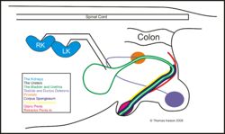
==Male== | ==Male== | ||
| − | + | [[Image:maleluttri.jpg|right|thumb|250px|<small><center>A schematic overview of the path of the male urethra</center></small>]] | |
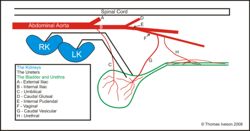
| + | [[Image:sumbsbladurtri.jpg|right|thumb|250px|<small><center>A schematic overview of the blood supply to the bladder and urethra</center></small>]] | ||
* Empties at tip of penis | * Empties at tip of penis | ||
* Divided into 3 parts | * Divided into 3 parts | ||
| Line 34: | Line 31: | ||
The '''urethralis''' muscle runs the entire length of the urethra and forms the external urethral sphincter. | The '''urethralis''' muscle runs the entire length of the urethra and forms the external urethral sphincter. | ||
| − | Unlike the [[Bladder - Anatomy & Physiology#Internal Urethral Sphincter|internal sphincter]] the external sphincter is composed of striated muscle fibres which are under the voluntary control of the somatic nervous system. | + | Unlike the [[Urinary Bladder - Anatomy & Physiology#Internal Urethral Sphincter|internal sphincter]] the external sphincter is composed of striated muscle fibres which are under the voluntary control of the somatic nervous system. |
=====Somatic Motor Supply - External Urethral Sphincter===== | =====Somatic Motor Supply - External Urethral Sphincter===== | ||
| Line 45: | Line 42: | ||
==Blood Supply== | ==Blood Supply== | ||
| − | Blood Supply comes from the Urethral Artery | + | Blood Supply comes from the Urethral Artery which is a branch of the vaginal artery which in turn is a branch of the internal pudendal which is a branch of the internal iliac. |
| + | |||
| + | ==Revision== | ||
| + | |||
| + | {{Template:Learning | ||
| + | |flashcards = Use the [[Urethra - Anatomy & Physiology - Renal Flash Cards - Anatomy & Physiology|flash card revision resource]] for this section to test yourself. | ||
| + | |dragster = [[Canine Urinary Radiographical Anatomy Resources (I & II)|Canine Male Urethrogram Radiographic Anatomy (II)]] | ||
| + | }} | ||
| + | |||
| + | |||
| + | ==Webinars== | ||
| + | <rss max="10" highlight="none">https://www.thewebinarvet.com/urogenital-and-reproduction/webinars/feed</rss> | ||
| + | |||
| + | [[Category:Lower Urinary Tract - Anatomy & Physiology]][[Category:Bullet Points]] | ||
Latest revision as of 16:05, 6 January 2023
Introduction
This muscular tube is the connection between the bladder and the external environment and plays a vital role in concious urinary continence.
The Layers of the Urethra
The layers of the walls of the urethra are largely similar to those of the urinary bladder apart from one significant difference; in both the male and the female the urethral submucosa has a network of veins which may contribute to continence by forming a kind of erectile tissue.
Female
- Empties at the external urethral orifice on the ventral wall of the vagina
- This is often at the vestibulo vaginal junction
- Only urine passes through it
- Length and diameter is species specific
- Short and wide - mare
- 2 fossa - bitch
- Suburethral diverticulum - cow and sow
Male
- Empties at tip of penis
- Divided into 3 parts
- Pre-prostatic - bladder neck to seminal hillock
- Prostatic portion - openings of deferant, vesicular and prostatic ducts
- Penile portion - ischial arch to penile tip
- Note the first 2 combined are called the pelvic portion
Muscles of the Urethra
The urethralis muscle runs the entire length of the urethra and forms the external urethral sphincter.
Unlike the internal sphincter the external sphincter is composed of striated muscle fibres which are under the voluntary control of the somatic nervous system.
Somatic Motor Supply - External Urethral Sphincter
- S1-S2
- Pudendal Nerve
- No synapse
- Innervates the urethral skeletal muscle - external urethral sphincter
- Function is to contract and retain urine
Blood Supply
Blood Supply comes from the Urethral Artery which is a branch of the vaginal artery which in turn is a branch of the internal pudendal which is a branch of the internal iliac.
Revision
| Urethra - Anatomy & Physiology Learning Resources | |
|---|---|
 Test your knowledge using drag and drop boxes |
Canine Male Urethrogram Radiographic Anatomy (II) |
 Test your knowledge using flashcard type questions |
Use the flash card revision resource for this section to test yourself. |
Webinars
Failed to load RSS feed from https://www.thewebinarvet.com/urogenital-and-reproduction/webinars/feed: Error parsing XML for RSS

