Difference between revisions of "Joints - Anatomy & Physiology"
Jump to navigation
Jump to search
m |
|||
| (19 intermediate revisions by 4 users not shown) | |||
| Line 1: | Line 1: | ||
| − | + | <big><center>[[Musculoskeletal System - Anatomy & Physiology|'''BACK TO MUSCULOSKELETAL ANATOMY AND PHYSIOLOGY''']]</center></big> | |
| + | |||
==Introduction== | ==Introduction== | ||
| − | + | Joints comprise broadly two categories: | |
| − | + | *'''Synarthroses''' form joints that are relatively rigid | |
| + | *'''Diarthroses''' form joints that are freely movable | ||
| + | Joint Function: | ||
| + | *Absorb force of impact, transfer force via cartilage to bone | ||
| + | *Allow a variable degree of movement | ||
| − | + | ==Fibrous Joints== | |
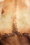
| + | [[Image: Skull sutures.jpg|thumb|right|100px|Sutures of the skull- Copyright Thegreenj]] | ||
| + | *Most occur in the skull: known as '''sutures''' | ||
| + | **Key in development: allow extension of individual bones during growth | ||
| + | **Gradually eliminated as ossification progresses | ||
| + | *'''Syndesmoses''': facing areas of two bones joined by connective tissue ligaments, very limited movement allowed | ||
| + | **Eg. Joints of the metacarpus in the horse | ||
| + | *'''Gomphosis''': attachment of tooth to bone within its socket | ||
| − | |||
| − | |||
| − | |||
| − | |||
| − | |||
| − | |||
| − | |||
| − | |||
| − | |||
| − | |||
| − | |||
| − | |||
| − | '''Symphysis''' | + | ==Cartilaginous Joints== |
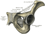
| − | + | [[Image:Grays Pelvic Symphysis.png|thumb|right|150px|Pelvic Symphysis, Gray's Anatomy - Wikimedia Commons 2008]] | |
| − | + | *'''Synchondroses''': eg. joints between epiphyses and diaphyses of juvenile long bones, disappear on maturity | |
| + | **Permanent synchondroses: the joint between the skull and '''hyoid''' | ||
| + | *'''Symphysis''': articulating bones are divided by a succession of tissues, with cartilage covering the bones or the tissue between | ||
| + | **Eg. mandibular, pelvic, vertebral | ||
| + | **Fibrocartilagenous joints | ||
| + | ***Form major union between vertebrae, except first two cervical vertebrae | ||
| + | ***''Nucleus pulposus'' is position eccentrically within ''annulus fibrosis'' | ||
| + | ***Vertebrae in thoracic region have '''conjugal ligaments''' | ||
| + | ****Extend from rib to rib on opposite sides | ||
| + | ****Strenghten the area over the discs | ||
==Synovial Joints== | ==Synovial Joints== | ||
| − | [[Image: | + | [[Image:Synovial Joint.jpg|thumb|right|150px|Synovial Joint - Wikimedia Commons 2008]] |
| − | Articulating joints are separated by a fluid-filled joint cavity, which is bounded by a synovial membrane | + | *Articulating joints are separated by a fluid-filled joint cavity, which is bounded by a synovial membrane |
| + | **Synovial membrane: pink connective tissue sheet, vascular and sensitive | ||
| + | ***Can be unsupported (membrane may pouch, allowing remote access), resting on an outer fibrous capsule, or separated from capsule by pads of fat | ||
| + | ***No continuous covering of cells | ||
| + | ***Where cells exist, they produce lubricant (aminoglycans) of synovial fluid | ||
| + | **Synovial fluid: Nourishes and lubricates [[Joints - normal#Articular cartilage|articular cartilage]] | ||
| + | ***Derived from synovial membrane cells and blood plasma | ||
| + | ***Normal amount in canine joint - 0.01 - 1.0 ml; possible in equine/bovine: 20-40mL | ||
| + | ***Transparent to light yellow (horses) | ||
| + | ***Usually very thick due to high hyaluronic acid, forms strands | ||
| + | ***Windrowing of cells on smear | ||
| + | ***Normal protein < 25g/l (all species) | ||
| + | ***Normal cell count: Large mononuclear cells, <12% neutrophils, <11% lymphocytes | ||
| + | ****Small animals - < 3 x 10e9/L | ||
| + | ****Horses - < 0.5 x 10e9/L | ||
| + | ****Cows - < 1 x 10e9/L | ||
| + | *Often the synovial membrane is reinforced by a fibrous capsule and ligaments restricting joint movement and providing stability | ||
| + | **Encloses bone and muscle insertions within joint capsule | ||
| + | **Supplied by blood vessels and nerve endings | ||
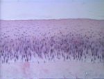
| + | *Articular cartilage covers the articular surfaces [[Image:Normal joint cartilage.jpg|right|thumb|150px|<small><center>Normal joint cartilage (Image courtesy Bristol Biomed Image Archive)</center></small>]] | ||
| + | **Usually, this is [[Bones and Cartilage - Anatomy & Physiology#Structure and Function of Cartilage|Hyaline]], although [[Bones and Cartilage - Anatomy & Physiology#Structure and Function of Cartilage|Fibrocartilage]] or fibrous tissue can substitute | ||
| + | **Articular cartilage is avascular and insensitive | ||
| + | ***Nutrients via diffusion from synovial fluid and nearby vessels (adjoining tissue and marrow cavities) | ||
| + | *Some joints possess intracapsular '''discs''' or '''menisci''' to provide congruence and enable complicated movements | ||
| + | **Eg. Temperomandibular joint, paired menisci of the stifle joint | ||
| + | **Limited response to injury, Little repair capacity | ||
| − | |||
| − | |||
| − | |||
| − | |||
| − | |||
| − | |||
| − | |||
| − | |||
| − | |||
| − | |||
==Joint Movements== | ==Joint Movements== | ||
| − | + | [[Image:Joint movements.jpg|thumb|right|150px|Flexion and Extension - Wikimedia Commons 2008]] | |
| − | + | *Translation: Flat surfaces slide against each other, producing no change in orientation of their bodies | |
| − | + | *Rotation: Moving bone turns on an axis perpendicular to articulation | |
| − | + | *Swing: Moving bone turns on an axis parallel to articulation | |
| − | + | *Flexion:(aka palmar flexion) Angle between two segments of a limb is reduced | |
| − | + | *Extension: Angle between two segments of a limb is increased | |
| − | + | *Overextension: (aka dorsal flexion) Eg, Posture of equine fetlock standing at rest | |
| − | + | *Adduction: Pendular movement toward the median plane | |
| − | + | *Abduction: Pendular movement away from the median plane | |
| − | + | *Circumduction: Combination of flexion and extension that allows a limb to create a circular movement | |
| − | |||
| − | |||
| − | |||
| − | |||
| − | |||
| − | |||
| − | |||
| − | |||
==Types of Joints== | ==Types of Joints== | ||
| − | + | *Plane Joint: describes translational movement; in reality, nonexistent, as all articular surfaces are curved | |
| − | + | *Hinge Joint: movement allowed in one plane only, inhibited by collateral ligaments and/or bony protuberances (eg. elbow joint) | |
| − | + | *Pivot Joint: comprises a peg fitted with a ring, movement occurs about the long axis of the peg (eg. radioulnar joint) | |
| − | + | *Condylar Joint: knuckle-shaped condyles vary in distance from one another allowing uniaxial movement with limited rotation (eg.femorotibial joint) | |
| − | + | *Ellipsoidal Joint: ovoid convex articulation allows movement in two planes at right angles with limited rotation (eg. radiocarpal joint) | |
| − | + | *Saddle Joint: also biaxial with a greater scope for rotation | |
| − | + | *Spheroidal Joint: (aka ball-and-socket) multiaxial movement allows for rotational movement in several planes (eg hip joint) | |
| − | |||
| − | |||
| − | |||
| − | |||
| − | |||
| − | |||
| − | |||
| − | |||
| − | |||
| − | |||
| − | |||
==Links== | ==Links== | ||
| + | *[[Joints - Pathology|Joint Pathology]] | ||
| − | + | <big><center>[[Musculoskeletal System - Anatomy & Physiology|'''BACK TO MUSCULOSKELETAL ANATOMY AND PHYSIOLOGY''']]</center></big> | |
| − | |||
| − | |||
| − | |||
| − | |||
Revision as of 19:37, 18 August 2008
Introduction
Joints comprise broadly two categories:
- Synarthroses form joints that are relatively rigid
- Diarthroses form joints that are freely movable
Joint Function:
- Absorb force of impact, transfer force via cartilage to bone
- Allow a variable degree of movement
Fibrous Joints
- Most occur in the skull: known as sutures
- Key in development: allow extension of individual bones during growth
- Gradually eliminated as ossification progresses
- Syndesmoses: facing areas of two bones joined by connective tissue ligaments, very limited movement allowed
- Eg. Joints of the metacarpus in the horse
- Gomphosis: attachment of tooth to bone within its socket
Cartilaginous Joints
- Synchondroses: eg. joints between epiphyses and diaphyses of juvenile long bones, disappear on maturity
- Permanent synchondroses: the joint between the skull and hyoid
- Symphysis: articulating bones are divided by a succession of tissues, with cartilage covering the bones or the tissue between
- Eg. mandibular, pelvic, vertebral
- Fibrocartilagenous joints
- Form major union between vertebrae, except first two cervical vertebrae
- Nucleus pulposus is position eccentrically within annulus fibrosis
- Vertebrae in thoracic region have conjugal ligaments
- Extend from rib to rib on opposite sides
- Strenghten the area over the discs
Synovial Joints
File:Synovial Joint.jpg
Synovial Joint - Wikimedia Commons 2008
- Articulating joints are separated by a fluid-filled joint cavity, which is bounded by a synovial membrane
- Synovial membrane: pink connective tissue sheet, vascular and sensitive
- Can be unsupported (membrane may pouch, allowing remote access), resting on an outer fibrous capsule, or separated from capsule by pads of fat
- No continuous covering of cells
- Where cells exist, they produce lubricant (aminoglycans) of synovial fluid
- Synovial fluid: Nourishes and lubricates articular cartilage
- Derived from synovial membrane cells and blood plasma
- Normal amount in canine joint - 0.01 - 1.0 ml; possible in equine/bovine: 20-40mL
- Transparent to light yellow (horses)
- Usually very thick due to high hyaluronic acid, forms strands
- Windrowing of cells on smear
- Normal protein < 25g/l (all species)
- Normal cell count: Large mononuclear cells, <12% neutrophils, <11% lymphocytes
- Small animals - < 3 x 10e9/L
- Horses - < 0.5 x 10e9/L
- Cows - < 1 x 10e9/L
- Synovial membrane: pink connective tissue sheet, vascular and sensitive
- Often the synovial membrane is reinforced by a fibrous capsule and ligaments restricting joint movement and providing stability
- Encloses bone and muscle insertions within joint capsule
- Supplied by blood vessels and nerve endings
- Articular cartilage covers the articular surfaces
- Usually, this is Hyaline, although Fibrocartilage or fibrous tissue can substitute
- Articular cartilage is avascular and insensitive
- Nutrients via diffusion from synovial fluid and nearby vessels (adjoining tissue and marrow cavities)
- Some joints possess intracapsular discs or menisci to provide congruence and enable complicated movements
- Eg. Temperomandibular joint, paired menisci of the stifle joint
- Limited response to injury, Little repair capacity
Joint Movements
File:Joint movements.jpg
Flexion and Extension - Wikimedia Commons 2008
- Translation: Flat surfaces slide against each other, producing no change in orientation of their bodies
- Rotation: Moving bone turns on an axis perpendicular to articulation
- Swing: Moving bone turns on an axis parallel to articulation
- Flexion:(aka palmar flexion) Angle between two segments of a limb is reduced
- Extension: Angle between two segments of a limb is increased
- Overextension: (aka dorsal flexion) Eg, Posture of equine fetlock standing at rest
- Adduction: Pendular movement toward the median plane
- Abduction: Pendular movement away from the median plane
- Circumduction: Combination of flexion and extension that allows a limb to create a circular movement
Types of Joints
- Plane Joint: describes translational movement; in reality, nonexistent, as all articular surfaces are curved
- Hinge Joint: movement allowed in one plane only, inhibited by collateral ligaments and/or bony protuberances (eg. elbow joint)
- Pivot Joint: comprises a peg fitted with a ring, movement occurs about the long axis of the peg (eg. radioulnar joint)
- Condylar Joint: knuckle-shaped condyles vary in distance from one another allowing uniaxial movement with limited rotation (eg.femorotibial joint)
- Ellipsoidal Joint: ovoid convex articulation allows movement in two planes at right angles with limited rotation (eg. radiocarpal joint)
- Saddle Joint: also biaxial with a greater scope for rotation
- Spheroidal Joint: (aka ball-and-socket) multiaxial movement allows for rotational movement in several planes (eg hip joint)


