Difference between revisions of "Renal Anatomy - Anatomy & Physiology"
| (43 intermediate revisions by 13 users not shown) | |||
| Line 1: | Line 1: | ||
| − | {{ | + | {{toplink |
| − | == | + | |backcolour = C1F0F6 |
| − | + | |linkpage =Urinary System - Anatomy & Physiology | |
| + | |linktext =URINARY SYSTEM | ||
| + | |maplink = Urinary System (Content Map) - Anatomy & Physiology | ||
| + | |pagetype =Anatomy | ||
| + | }} | ||
| + | <br> | ||
| + | {|align="right" | ||
| + | |__TOC__ | ||
| + | |} | ||
| + | ==Common Anatomy== | ||
| − | |||
| − | |||
| − | |||
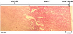
[[Image:basicnormkidap.jpg|right|thumb|250px|<small><center>Histology section of a normal kidney (© RVC 2008)</center></small>]] | [[Image:basicnormkidap.jpg|right|thumb|250px|<small><center>Histology section of a normal kidney (© RVC 2008)</center></small>]] | ||
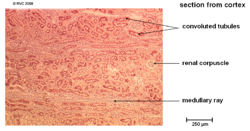
[[Image:normkidcortap.jpg|right|thumb|250px|<small><center>Histology section of a normal renal cortex (© RVC 2008)</center></small>]] | [[Image:normkidcortap.jpg|right|thumb|250px|<small><center>Histology section of a normal renal cortex (© RVC 2008)</center></small>]] | ||
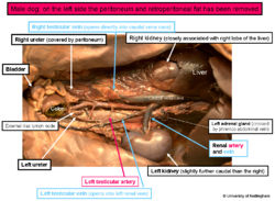
[[Image:promaledogab.jpg|right|thumb|250px|<small><center>A prosection of the abdomen of a male dog (© UoN 2008)</center></small>]] | [[Image:promaledogab.jpg|right|thumb|250px|<small><center>A prosection of the abdomen of a male dog (© UoN 2008)</center></small>]] | ||
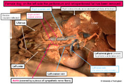
[[Image:profemaledogab.jpg|right|thumb|250px|<small><center>A prosection of the abdomen of a female dog(© UoN 2008)</center></small>]] | [[Image:profemaledogab.jpg|right|thumb|250px|<small><center>A prosection of the abdomen of a female dog(© UoN 2008)</center></small>]] | ||
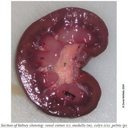
| + | [[Image:sagkidlabelled.jpg|right|thumb|250px|<small><center>A labelled saggital section of a lamb kidney(Courtesy of Donal McNally - University of Nottingham)</center></small>]] | ||
* The kidney is the part of the urinary tract where blood is filtered and urine is produced. | * The kidney is the part of the urinary tract where blood is filtered and urine is produced. | ||
* The kidneys are paired and lie in a retroperitoneal position. | * The kidneys are paired and lie in a retroperitoneal position. | ||
* They are positioned in the caudo-dorsal abdomen. | * They are positioned in the caudo-dorsal abdomen. | ||
* They lie within a splitting of the sublumbar fascia. This also often contains a large quantity of fat to cushion and protect the kidneys from the pressure of other organs | * They lie within a splitting of the sublumbar fascia. This also often contains a large quantity of fat to cushion and protect the kidneys from the pressure of other organs | ||
| − | * The right kidney is most cranial in all species except the pig | + | * The right kidney is most cranial in all species except the pig. |
* In species where the right kidney is most cranial it lies in a small fossa of the caudate liver lobe. | * In species where the right kidney is most cranial it lies in a small fossa of the caudate liver lobe. | ||
* However the left kidney is the most mobile. | * However the left kidney is the most mobile. | ||
| Line 29: | Line 36: | ||
* External zone | * External zone | ||
* Internal zone (juxtamedullar) | * Internal zone (juxtamedullar) | ||
| − | * Contains the following parts of the [[ | + | * Contains the following parts of the [[The Nephron - Anatomy & Physiology| nephron]] |
| − | ** [[Nephron | + | ** [[Microscopic Anatomy of the Nephron - Anatomy & Physiology#The Renal Corpuscle| Renal corpuscle]] |
| − | ** [[Nephron | + | ** [[Microscopic Anatomy of the Nephron - Anatomy & Physiology#Proximal Tubule| Proximal convoluted tubule]] |
| − | ** [[Nephron | + | ** [[Microscopic Anatomy of the Nephron - Anatomy & Physiology#The Distal Tubule| Distal convoluted tubule]] |
| − | ** [[ | + | ** [[Useful definitions - Renal Anatomy & Physiology#M| Medullary Rays]] |
===Renal Medulla=== | ===Renal Medulla=== | ||
| − | |||
* Contains medullary pyramids | * Contains medullary pyramids | ||
* The part nearest the cortex is the base of the pyramid which narrows to form the inner part - renal papilla | * The part nearest the cortex is the base of the pyramid which narrows to form the inner part - renal papilla | ||
* The medulla can be split into two parts, the outer and the inner | * The medulla can be split into two parts, the outer and the inner | ||
| − | * Different parts of the [[ | + | * Different parts of the [[The Nephron - Anatomy & Physiology| nephron]] reside in these areas |
| − | + | * The outer medulla, which can be further divided into the outer and inner stripe, contains the following parts of the [[The Nephron - Anatomy & Physiology| nephron]]: | |
| − | The outer medulla can be further divided into the outer and inner stripe | + | ** [[Microscopic Anatomy of the Nephron - Anatomy & Physiology#Proximal Tubule| Proximal straight tubules]] |
| − | + | ** [[Microscopic Anatomy of the Nephron - Anatomy & Physiology#The Distal Tubule| Distal straight tubule]] | |
| − | + | ** [[Microscopic Anatomy of the Nephron - Anatomy & Physiology#The Loop of Henle| Loop of henle]] | |
| − | + | ** [[Microscopic Anatomy of the Nephron - Anatomy & Physiology#Collecting Duct| Collecting ducts]] | |
| − | * [[Nephron | + | ** [[Microscopic Anatomy of the Nephron - Anatomy & Physiology #The Vasa Recta| Vasa recta]] |
| − | * [[Nephron | ||
| − | * [[Nephron | ||
| − | * [[Nephron | ||
| − | * [[Nephron | ||
| − | |||
| − | |||
| − | |||
| − | |||
| − | |||
| − | |||
| − | |||
| − | |||
| − | |||
| − | |||
| − | |||
* The inner medulla however only contains the following parts: | * The inner medulla however only contains the following parts: | ||
| − | ** [[Nephron | + | ** [[Microscopic Anatomy of the Nephron - Anatomy & Physiology#The Loop of Henle| Loop of henle]] |
| − | ** [[Nephron | + | ** [[Microscopic Anatomy of the Nephron - Anatomy & Physiology#Collecting Duct| Collecting ducts]] |
| − | ** [[Nephron | + | ** [[Microscopic Anatomy of the Nephron - Anatomy & Physiology #The Vasa Recta| Vasa recta]] |
===Renal Pelvis=== | ===Renal Pelvis=== | ||
| Line 72: | Line 63: | ||
* All papillary ducts open here | * All papillary ducts open here | ||
* The renal pelvis then drains into the ureters | * The renal pelvis then drains into the ureters | ||
| − | |||
* Varies between species | * Varies between species | ||
* Absent in cow | * Absent in cow | ||
* Contains mucous glands in the horse | * Contains mucous glands in the horse | ||
| − | + | * The kidney is divided into [[Useful definitions - Renal Anatomy & Physiology | renal lobes]] from a structural point of view. | |
| − | + | * These are then subdivided into [[The Nephron - Anatomy & Physiology| nephrons]], [[Useful definitions - Renal Anatomy & Physiology | a medullary ray]] and [[Microscopic Anatomy of the Nephron - Anatomy & Physiology#Collecting Duct| a collecting duct]] to form a [[Useful definitions - Renal Anatomy & Physiology | renal lobule]] | |
| − | * The kidney is divided into [[ | ||
| − | * These | ||
| − | |||
| − | |||
| − | |||
| − | |||
| − | |||
===Innervation=== | ===Innervation=== | ||
| Line 104: | Line 87: | ||
These species all have similar renal anatomy. Their kidneys are relatively short and thick and they are the traditional kidney bean shape. They have a smooth outer surface and have a single renal papilla. The renal pelvis is large and irregular with recesses which are finger like processes. | These species all have similar renal anatomy. Their kidneys are relatively short and thick and they are the traditional kidney bean shape. They have a smooth outer surface and have a single renal papilla. The renal pelvis is large and irregular with recesses which are finger like processes. | ||
| − | The kidney of the feline is relatively bigger than the other species and is quite distinctive because the sub-capsular veins which run towards the hilum are visible | + | The kidney of the feline is relatively bigger than the other species and is quite distinctive because the sub-capsular veins which run towards the hilum are visible. |
===Bovine=== | ===Bovine=== | ||
| − | + | The kidneys of the bovine do not lose their foetal lobulation. In fact the surface of each kidney is divided into approximately 12 lobules. The right kidney is flattened and ellipsoidal where as the left kidney is thicker at the caudal end than the cranial. Each kidney is surrounded by the capsula adiposa; a layer of fat. Despite what it’s externally lobulated appearance may suggest the cortex of the bovine kidney is continuous and the kidney is of multipyramidal type. The bovine kidney has no renal pelvis but rather the [[Ureters - Anatomy & Physiology | Ureters]] enters the kidney and divide into a cranial and caudal branch. These branches then subdivide and the papilla at the apex of the pyramids open and drain into these. | |
| − | The kidneys of the bovine do not lose their foetal lobulation. In fact the surface of each kidney is divided into approximately 12 lobules. The right kidney is flattened and ellipsoidal where as the left kidney is thicker at the caudal end than the cranial. Each kidney is surrounded by the capsula adiposa; a layer of fat. Despite what it’s externally lobulated appearance may suggest | ||
The right ureter leaves the kidney and passes along the roof of the abdomen to the pelvis in a fairly standard pattern. The left ureter however moves across the dorsal surface of its kidney to return to the midline and follow a course as if the kidney was located on the left. (both kidneys in the bovine are located on the right see the anatomical landmarks section for further details) | The right ureter leaves the kidney and passes along the roof of the abdomen to the pelvis in a fairly standard pattern. The left ureter however moves across the dorsal surface of its kidney to return to the midline and follow a course as if the kidney was located on the left. (both kidneys in the bovine are located on the right see the anatomical landmarks section for further details) | ||
===Porcine=== | ===Porcine=== | ||
| − | + | The kidneys are dorsoventrally flattened. The renal pelvis opens into quite a large space of two major calicyes from which bud about 10 minor calyces. These attach to one renal papillae each. The kidneys have a smooth surface. | |
| − | The kidneys are dorsoventrally flattened. The renal pelvis opens into quite a large space of two major | ||
===Equine=== | ===Equine=== | ||
| Line 130: | Line 111: | ||
Medially to the right kidney you will find the caudal vena cava and the right adrenal gland can be found dorsolateral to this structure but still medially to the kidney. Ventrally can be found the Descending [[Duodenum - Anatomy & Physiology|duodenum]] with the right pancreatic limb more towards the ventromedial aspect. In females the right ovary can be found caudoventrally. | Medially to the right kidney you will find the caudal vena cava and the right adrenal gland can be found dorsolateral to this structure but still medially to the kidney. Ventrally can be found the Descending [[Duodenum - Anatomy & Physiology|duodenum]] with the right pancreatic limb more towards the ventromedial aspect. In females the right ovary can be found caudoventrally. | ||
| − | The '''Cranial pole''' of the left kidney contacts the [[ | + | The '''Cranial pole''' of the left kidney contacts the [[Stomach and Abomasum - Anatomy & Physiology | Greater curvature of stomach]] it also contacts the [[Spleen - Anatomy & Physiology|spleen]] on its dorsomedial aspect as well as potentially contacting the left limb of the pancreas. The left adrenal gland can be found at the medial aspect of the cranial pole. The '''Caudal pole''' of the left kidney contacts the [[Small Intestine - Anatomy & Physiology|Small intestine]] and the Descending [[Colon - Anatomy & Physiology|colon]]. At its caudoventral aspect in females is found the left Ovary. |
===Ruminants=== | ===Ruminants=== | ||
| Line 184: | Line 165: | ||
===Internal Vascularisation=== | ===Internal Vascularisation=== | ||
| − | Once the renal artery enters the kidney is divides into the interlobar arteries. These pass through the gaps between the renal pyramids as the reach the junctions between the cortex and medullar they branch into the arcuate arteries which move over the base of the pyramids. From these come the interlobular arteries which supply the individual lobules of the cortex. These arteries then branch many times to supply individual glomeruli. The capillaries of the glomerulus then rejoin to form one vessel which then forms the [[ | + | Once the renal artery enters the kidney is divides into the interlobar arteries. These pass through the gaps between the renal pyramids as the reach the junctions between the cortex and medullar they branch into the arcuate arteries which move over the base of the pyramids. From these come the interlobular arteries which supply the individual lobules of the cortex. These arteries then branch many times to supply individual glomeruli. The capillaries of the glomerulus then rejoin to form one vessel which then forms the [[Peritubular Capillaries - Anatomy & Physiology|peritubular capillaries]] of that nephron. The interlobular arteries are examples of end arterioles and there are few anastomoses. Obstruction of one of these arterioles causes ischaemic damage in the kidneys. This is also potentially the case with the interlobar arteries. |
| + | |||
| − | |||
| − | |||
| − | |||
| − | |||
| − | |||
| − | |||
| − | == | + | ==Renal Pathology== |
| − | + | The pathology of the kidney is beyond the scope of this section but can be found [[Urinary System - Pathology| here]] | |
| − | [[ | ||
Revision as of 17:11, 7 September 2008
|
|
Common Anatomy
- The kidney is the part of the urinary tract where blood is filtered and urine is produced.
- The kidneys are paired and lie in a retroperitoneal position.
- They are positioned in the caudo-dorsal abdomen.
- They lie within a splitting of the sublumbar fascia. This also often contains a large quantity of fat to cushion and protect the kidneys from the pressure of other organs
- The right kidney is most cranial in all species except the pig.
- In species where the right kidney is most cranial it lies in a small fossa of the caudate liver lobe.
- However the left kidney is the most mobile.
- During development all species begin with a multi-lobed structure but a varying degree of fusion occurs between the species giving rise to the various different characteristics seen.
The Basic Components of the Kidney
Outer fibrous capsule
A tough outer capsule surrounds the parenchyma and this prevents the kidney expanding. It is easily stripped away from a healthy kidney but adheres where pathology has occured.
Renal Cortex
- 2 Parts
- External zone
- Internal zone (juxtamedullar)
- Contains the following parts of the nephron
Renal Medulla
- Contains medullary pyramids
- The part nearest the cortex is the base of the pyramid which narrows to form the inner part - renal papilla
- The medulla can be split into two parts, the outer and the inner
- Different parts of the nephron reside in these areas
- The outer medulla, which can be further divided into the outer and inner stripe, contains the following parts of the nephron:
- The inner medulla however only contains the following parts:
Renal Pelvis
- The renal sinus is located within an indentation on the medial side of the kidney
- The renal pelvis is located within the renal sinus
- All papillary ducts open here
- The renal pelvis then drains into the ureters
- Varies between species
- Absent in cow
- Contains mucous glands in the horse
- The kidney is divided into renal lobes from a structural point of view.
- These are then subdivided into nephrons, a medullary ray and a collecting duct to form a renal lobule
Innervation
- Sympathetic and parasympathetic fibres from solar plexus
- Travel with renal arteries
- Sympathetic fibres synapse in coeliac ganglion and cranial mesenteric ganglion
Lymphatic Drainage
Renal lymph nodes
Anatomical Species Differences
The various species have major and rather striking differences in the structure of their kidneys.
Canine, Feline, and Ovine
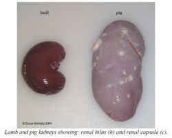
These species all have similar renal anatomy. Their kidneys are relatively short and thick and they are the traditional kidney bean shape. They have a smooth outer surface and have a single renal papilla. The renal pelvis is large and irregular with recesses which are finger like processes.
The kidney of the feline is relatively bigger than the other species and is quite distinctive because the sub-capsular veins which run towards the hilum are visible.
Bovine
The kidneys of the bovine do not lose their foetal lobulation. In fact the surface of each kidney is divided into approximately 12 lobules. The right kidney is flattened and ellipsoidal where as the left kidney is thicker at the caudal end than the cranial. Each kidney is surrounded by the capsula adiposa; a layer of fat. Despite what it’s externally lobulated appearance may suggest the cortex of the bovine kidney is continuous and the kidney is of multipyramidal type. The bovine kidney has no renal pelvis but rather the Ureters enters the kidney and divide into a cranial and caudal branch. These branches then subdivide and the papilla at the apex of the pyramids open and drain into these.
The right ureter leaves the kidney and passes along the roof of the abdomen to the pelvis in a fairly standard pattern. The left ureter however moves across the dorsal surface of its kidney to return to the midline and follow a course as if the kidney was located on the left. (both kidneys in the bovine are located on the right see the anatomical landmarks section for further details)
Porcine
The kidneys are dorsoventrally flattened. The renal pelvis opens into quite a large space of two major calicyes from which bud about 10 minor calyces. These attach to one renal papillae each. The kidneys have a smooth surface.
Equine
The equine kidneys not only have very different shapes compared to the rest of the domestic species but they also each have a different shape. The right kidney is described as heart shaped whilst the left is described as being pyramidal. Each organ weighs approximately 700g and both are dorsoventrally flattened. The kidneys are basically unipyramidal and the only demarcation between what were the multiple pyramids of the foetus are the interlobar arteries. This is not always the case in the foal where it is common to be able to identify lobes and the external surface is not always smooth. The horse has a single renal papilla like the dog and its renal pelvis is large and irregular with 2 recesses (finger like processes). The cells of its pelvis secret mucin giving the urine its cloudy appearance.
Anatomical Landmarks
Each species has a slightly different orientation of the kidneys within the abdomen.
Carnivores
The kidneys in these species are very mobile. Especially the left one and especially in the feline. These anatomical landmarks describe the most common locations. The right kidney is more cranial than the left and tends to lie beneath L1 - L3 where as the left tends to lie under L2-L4. The reason the right kidney is less mobile than the left is that it tends to be almost entirely enclosed within the renal fossa of the caudate lobe of the liver. The kidneys are enclosed within a layer of fat and this increases with the obesity of the animal.
Medially to the right kidney you will find the caudal vena cava and the right adrenal gland can be found dorsolateral to this structure but still medially to the kidney. Ventrally can be found the Descending duodenum with the right pancreatic limb more towards the ventromedial aspect. In females the right ovary can be found caudoventrally.
The Cranial pole of the left kidney contacts the Greater curvature of stomach it also contacts the spleen on its dorsomedial aspect as well as potentially contacting the left limb of the pancreas. The left adrenal gland can be found at the medial aspect of the cranial pole. The Caudal pole of the left kidney contacts the Small intestine and the Descending colon. At its caudoventral aspect in females is found the left Ovary.
Ruminants
Due to the rumen taking up most of the left side of the abdomen it is normal in the bovine to find both of the kidneys on the right side. In the ovine the kidneys are surrounded by very thick masses of fat which reduce the impact of the rumen on their location. The kidneys in the living animal vary their position substantially with respiration and with the pressure of the other organs.
The right kidney has a retroperitoneal attachment to the "sublumbar" musculature. Cranially it touches the liver. In the dead specimen the right kidney occupies the region between the last rib and somewhere between the 2nd and 3rd lumbar vertebrae’s transverse process. The right kidney usually contacts pancreas, duodenum, adrenal glands and colon as well as the afore mentioned liver.
The left kidney in the bovine is to be found caudoventrally to the right one usually in the region between the 2nd and 4th lumbar vertebrae
Porcine
The left and right kidneys are more or less aligned in the pig unlike other species. The kidneys of the pig are embedded in lots of fat and lie against the psoas muscle. They span the distance between the last rib cranially and the 4th lumber vertebrae caudally. The right kidney does not touch the liver unlike other species and is related ventrally to the descending duodenum and jejunum. The left kidney is ventrally related to the ascending colon, the base of the caecum and the pancreas.
Equine
The kidneys of the horse are both enclosed in a fat capsule. Dorsally they rest against the psoas muscle and against the diaphragm.
The right kidney is to be found ventrally to and between the last 2 ribs and first lumbar transverse process. Cranially it touches the liver and caudally it is attached to the pancreas and the base of caecum. The duodenum winds around its lateral and then ventral surfaces. Medially is the caudal vena cava and adrenal gland.
The left kidney is between the last rib and 3rd transverse process. Its ventral surface is almost completely covered by the peritoneum and contacts the small intestine and and small colon. The spleen contacts it cranioventrally. Medially is the left adrenal gland and aorta.
Renal Blood Supply
- Supplied by renal arteries
- Arise from aorta
- 3-4mm diameter - dog
- Often divide into dorsal and ventral branches before entering kidney
- Common to find double the normal number - dog
Left Renal Vessels
- Left renal artery
- Originates 2cm caudal to the right renal artery
- ~3-4cm long - dog
- Left renal vein
- Immediately ventral to artery
- ~3-4cm long - dog
Right Renal Vessels
- Right Renal Artery
- 2cm cranial to left renal artery
- 4 cm caudal to cranial mesenteric artery
- Passes dorsal over the caudal vena cava
- ~4-5cm long - dog
- Right Renal Vein
- Ventral to artery
- ~4-5cm long - dog
Internal Vascularisation
Once the renal artery enters the kidney is divides into the interlobar arteries. These pass through the gaps between the renal pyramids as the reach the junctions between the cortex and medullar they branch into the arcuate arteries which move over the base of the pyramids. From these come the interlobular arteries which supply the individual lobules of the cortex. These arteries then branch many times to supply individual glomeruli. The capillaries of the glomerulus then rejoin to form one vessel which then forms the peritubular capillaries of that nephron. The interlobular arteries are examples of end arterioles and there are few anastomoses. Obstruction of one of these arterioles causes ischaemic damage in the kidneys. This is also potentially the case with the interlobar arteries.
Renal Pathology
The pathology of the kidney is beyond the scope of this section but can be found here
