Difference between revisions of "Nephron - Anatomy & Physiology"
Jump to navigation
Jump to search
| (22 intermediate revisions by 3 users not shown) | |||
| Line 1: | Line 1: | ||
| + | {{toplink | ||
| + | |backcolour = C1F0F6 | ||
| + | |linkpage =Urinary System (Table) - Anatomy & Physiology | ||
| + | |linktext =URINARY SYSTEM | ||
| + | |thispagetable = The Nephron (Table) - Anatomy & Physiology | ||
| + | |thispagemap = Urinary System (Content Map) - Anatomy & Physiology | ||
| + | |pagetype =Anatomy | ||
| + | }} | ||
| + | <br> | ||
| + | <!--The Nephron - Anatomy & Physiology Background--> | ||
| + | {{infotable | ||
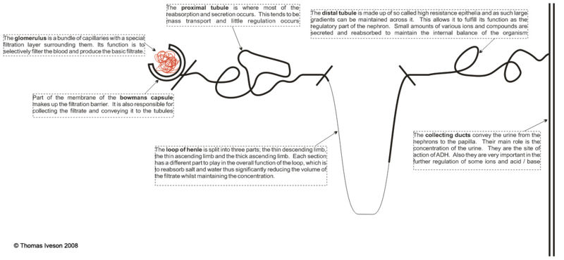
| + | The nephron of the kidney is made up of two major parts; the renal corpuscle and the tubules. These are then both sub-divided into various parts and overall it is this structure which allows the kidney to filter the blood and then alter the composition of this filtrate to ensure that waste products are excreted and useful compounds preserved. | ||
| + | |||
| + | The renal corpuscle can be subdivided into the glomerulus and the bowmans capsule. The tubules are split into the proximal tubule, the loop of henle, the distal tubule and the collecting ducts. | ||
| + | |||
| + | <center>[[Image:nephovertri.jpg|800px]]</center> | ||
| + | }} | ||
| + | |||
<!--The Nephron - Anatomy & Physiology--> | <!--The Nephron - Anatomy & Physiology--> | ||
{{infotable | {{infotable | ||
|Maintitle = The Nephron - Anatomy & Physiology | |Maintitle = The Nephron - Anatomy & Physiology | ||
|Maintitlebackcolour = 66CC33 | |Maintitlebackcolour = 66CC33 | ||
| − | |||
| − | |||
| − | |||
| − | |||
| − | |||
|subheading1colour = 66ff33 | |subheading1colour = 66ff33 | ||
| − | |subheading1 = [[ | + | |subheading1 = [[Macroscopic Renal Anatomy - Anatomy & Physiology|Macroscopic Renal Anatomy]] |
|subheading1width =25 | |subheading1width =25 | ||
| − | |subheading1text = <center>[[ | + | |subheading1text = <center>[[Macroscopic Renal Anatomy - Anatomy & Physiology#Common Anatomy|Common Anatomy]], [[Macroscopic Renal Anatomy - Anatomy & Physiology#Anatomical Species Differences|Anatomical Species Differences]], [[Macroscopic Renal Anatomy - Anatomy & Physiology#Anatomical Landmarks|Anatomical Landmarks]]</center> |
|subheading2colour = 66ff33 | |subheading2colour = 66ff33 | ||
| − | |subheading2 = [[ | + | |subheading2 = [[The Nephron (Table) - Anatomy & Physiology|The Nephron]] |
|subheading2width =25 | |subheading2width =25 | ||
|subheading2text = <center></center> | |subheading2text = <center></center> | ||
|subheading3colour = 66ff33 | |subheading3colour = 66ff33 | ||
| − | |subheading3 = [[ | + | |subheading3 = [[Kidney - Blood Pressure - Physiology|Blood Pressure]] |
|subheading3width =25 | |subheading3width =25 | ||
|subheading3text = <center></Center> | |subheading3text = <center></Center> | ||
|subheading4colour = 66ff33 | |subheading4colour = 66ff33 | ||
| − | |subheading4 = [[ | + | |subheading4 = [[The Endocrine Function of the Kidney - Anatomy & Physiology|The Endocrine Function of the Kidney]] |
|subheading4width =25 | |subheading4width =25 | ||
|subheading4text = <center></Center> | |subheading4text = <center></Center> | ||
}} | }} | ||
| − | |||
| − | |||
| − | |||
| − | |||
| − | |||
| − | |||
| − | |||
Revision as of 17:55, 9 September 2008
|
|
{{infotable
The nephron of the kidney is made up of two major parts; the renal corpuscle and the tubules. These are then both sub-divided into various parts and overall it is this structure which allows the kidney to filter the blood and then alter the composition of this filtrate to ensure that waste products are excreted and useful compounds preserved.
The renal corpuscle can be subdivided into the glomerulus and the bowmans capsule. The tubules are split into the proximal tubule, the loop of henle, the distal tubule and the collecting ducts.

}}
The Nephron - Anatomy & Physiology |
|---|
Macroscopic Renal Anatomy |
The Nephron |
Blood Pressure |
The Endocrine Function of the Kidney |
|---|---|---|---|