Difference between revisions of "Gastric Dilatation and Volvulus"
| Line 36: | Line 36: | ||
===Haematology=== | ===Haematology=== | ||
| + | *Increased haematocrit | ||
| + | *DIC (thrombocytopaenia, increased firbin degradation products, prolonged patial thromboplastin time and reduced antithrombin III. | ||
===Biochemistry=== | ===Biochemistry=== | ||
| + | Most commonly find hypokalaemia and metabolic acidosis. The acidosis is caused hypoperfusion and anaerobic metabolism leading to lactic acid accumulation. Respiratory acidosis and alkalosis may also be present due to hypo- and hyperventilation. | ||
===Diagnostic imaging=== | ===Diagnostic imaging=== | ||
| + | Best performed after fluid therapy and gastric decompression. It allows distinction between GD and GDV: | ||
| + | *Gastric dilatation: gas distension, on Right Lateral shows air in the fundus. | ||
| + | *Gastric dilatation and volvulus: pylorus moves dorsally and left with a compartmentalized stomach. | ||
| + | A right lateral view will show a large fundus ventrally, with a smaller gas filled pylorus located dorsally to that. These are seperated by a soft tissue strip. The contrast of the abdomen may be lost indicating peritonitis or haemabdomen. Gastric rupture would show as pneumoperitoneum and increased contrast. | ||
==Treatment== | ==Treatment== | ||
The most important first line treatments are [[Principles of Fluid Therapy|fluid therapy]] and gastric decompression | The most important first line treatments are [[Principles of Fluid Therapy|fluid therapy]] and gastric decompression | ||
===Fluid therapy=== | ===Fluid therapy=== | ||
| + | Should be individualised to the patient due to the varying nature of the acid-base disturbances. | ||
===Gastric decompression=== | ===Gastric decompression=== | ||
Revision as of 08:39, 21 August 2009
| This article is still under construction. |
Signalment
- Large deep chested breeds including:
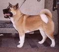
Akitas
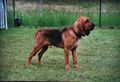
Bloodhounds
Collies
Great Danes
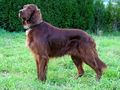
Irish Setters
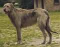
Wolfhounds
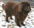
Newfoundlands
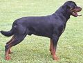
Rottweilers
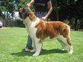
Saint Bernards

Standard Poodles
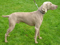
Weirmaraners


Description
Gastric dilatation (GD) and Gastric dilatation and volvulus (GDV) are caused by the stomach distending with air. In GDV the stomach twists around its axis with both conditions leading to compression of the caudal vena cava. GDV can lead to hypovolaemic shock, splenic torsion, gastric wall ischaemia, abdominal viscera congestion, endotoxic shock and disseminated intravascular coagulation (DIC)
Diagnosis
History and Clinical signs
- Abdominal distension
- Non-productive retching
- Weakness
- Collapse
- Salivation
- Abdominal tympany
- Tachycardia
- Pallor
- Hypothermia
- Cardiac arrythmias (ventricular premature beats, ventricular tachycardia)
Haematology
- Increased haematocrit
- DIC (thrombocytopaenia, increased firbin degradation products, prolonged patial thromboplastin time and reduced antithrombin III.
Biochemistry
Most commonly find hypokalaemia and metabolic acidosis. The acidosis is caused hypoperfusion and anaerobic metabolism leading to lactic acid accumulation. Respiratory acidosis and alkalosis may also be present due to hypo- and hyperventilation.
Diagnostic imaging
Best performed after fluid therapy and gastric decompression. It allows distinction between GD and GDV:
- Gastric dilatation: gas distension, on Right Lateral shows air in the fundus.
- Gastric dilatation and volvulus: pylorus moves dorsally and left with a compartmentalized stomach.
A right lateral view will show a large fundus ventrally, with a smaller gas filled pylorus located dorsally to that. These are seperated by a soft tissue strip. The contrast of the abdomen may be lost indicating peritonitis or haemabdomen. Gastric rupture would show as pneumoperitoneum and increased contrast.
Treatment
The most important first line treatments are fluid therapy and gastric decompression
Fluid therapy
Should be individualised to the patient due to the varying nature of the acid-base disturbances.