Difference between revisions of "Coccidia"
Jump to navigation
Jump to search
(Redirected page to Category:Coccidia) |
|||
| (11 intermediate revisions by the same user not shown) | |||
| Line 1: | Line 1: | ||
| − | # | + | ==Introduction== |
| + | |||
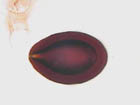
| + | [[Image:Coccidia.jpg|thumb|right|150px|Coccidia - Joel Mills]] | ||
| + | |||
| + | *The '''oocyst''' is the resistant stage in the environment | ||
| + | |||
| + | *The infective '''sporozoite''' is released from the oocyst | ||
| + | |||
| + | *Inside the host, the sporozoites invade the intestinal epithelial tissue | ||
| + | **Sporozoites feed and grow | ||
| + | |||
| + | *As the sporozoite grows the nucleus divides forming a '''schizont''' | ||
| + | |||
| + | *The schizont contains numerous elongated '''merozoites''' | ||
| + | |||
| + | *The formation of merozoites is the first asexual reproductive stage called '''schizogony''' | ||
| + | |||
| + | *The schizont ruptures releasing the merozoites which also invade the epithelial cells | ||
| + | |||
| + | *Another generation of schizonts form which is the beginning of the sexual phase of reproduction called '''gametogony''' | ||
| + | |||
| + | *The merozoites form male '''microgamonts''' or female '''macrogamonts''' | ||
| + | **Collectively known as gamonts or gametocytes | ||
| + | |||
| + | *The microgamonts released from the microgametocyte penetrate and fertilise the macrogamont (which is contained within the macrogametocyte) | ||
| + | |||
| + | *Gametogony forms the '''zygote''' | ||
| + | **Surrounded by a cyst wall | ||
| + | **Forms the '''oocyst''' | ||
| + | |||
| + | *The oocyst is passed in the faeces and is unsporulated | ||
| + | |||
| + | *The oocyst becomes sporulated in the second asexual reproductive phase called '''sporogony''' | ||
| + | |||
| + | *Once the oocyst is sporulated it is infective | ||
| + | |||
| + | <big> | ||
| + | '''[[Eimeria spp.|''Eimeria'' spp.]] | ||
| + | |||
| + | '''[[Isospora spp.|''Isospora'' spp.]] | ||
| + | |||
| + | '''[[Coccidiosis]] | ||
| + | |||
| + | '''[[Coccidia - Poultry|Coccidia of Poultry]] | ||
| + | |||
| + | '''[[Coccidiosis - Poultry]] | ||
| + | |||
| + | '''[[Coccidiosis - Turkey|Coccidia of Turkeys]] | ||
| + | |||
| + | '''[[Coccidiosis - Geese|Coccidia of Geese]] | ||
| + | |||
| + | '''[[Coccidiosis - Duck|Coccidia of Ducks]] | ||
| + | |||
| + | '''[[Coccidiosis - Game Birds|Coccidia of Game Birds]] | ||
| + | |||
| + | |||
| + | ==Coccidia of Cattle== | ||
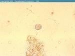
| + | [[Image:Coccidia ruminant.jpg|thumb|right|150px|''Eimeria'' sp. of ruminants - Joaquim Castellà Veterinary Parasitology Universitat Autònoma de Barcelona]] | ||
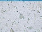
| + | [[Image:Coccidia oocyst ruminant.jpg|thumb|right|150px|Coccidia oocyst from ruminant faeces - Joaquim Castellà Veterinary Parasitology Universitat Autònoma de Barcelona]] | ||
| + | *Many species affect cattle | ||
| + | |||
| + | *Cattle under a year old are usually infected sporadically | ||
| + | |||
| + | *2-3 week prepatent period | ||
| + | |||
| + | *''Eimeria bovis'' | ||
| + | **Endogenous stages in central lacteal of villi and epithelial cells of [[Caecum - Anatomy & Physiology|caecum]] and [[Colon - Anatomy & Physiology|colon]] | ||
| + | **Causes [[Intestine Diarrhoea - Pathology|diarrhoea]] and enteritis | ||
| + | **Oocysts are 28x20μm | ||
| + | **Moderately pathogenic | ||
| + | |||
| + | *''Eimeria zuernii'' | ||
| + | **Endogenous stages in connective tissue of lamina propria of the lower [[Small Intestine - Anatomy & Physiology|small intestine]] and in the epithelial cells of the [[Caecum - Anatomy & Physiology|caecum]] and [[Colon - Anatomy & Physiology|colon]] | ||
| + | **More pathogenic than ''Eimeria bovis'' | ||
| + | **Causes blood stained dysentery, tenesmus and sloughed mucosa | ||
| + | **Oocysts are spherical and measure 16μm | ||
| + | |||
| + | *Mainly occurs in calves in poor conditions and bought-in calves | ||
| + | **Also occurs in suckler calves turned out in spring | ||
| + | |||
| + | *''Eimeria alabamensis'' associated with [[Intestine Diarrhoea - Pathology|diarrhoea]] in calves after spring turnout | ||
| + | |||
| + | *[[Materno-fetal immunity - WikiBlood#Passive transfer via colostrum|Passive immunity]] is sufficient during the neonatal period | ||
| + | |||
| + | *Can be concurrent with cryptosporidium, viral and bacterial agents | ||
| + | |||
| + | '''Diagnosis''' | ||
| + | *History, clinical signs, [[Intestine Diarrhoea - Pathology|diarrhoea]] (often with blood) and a decrease in weight gain | ||
| + | |||
| + | *Post-mortem | ||
| + | **Diffuse inflammation and thickening of [[Caecum - Anatomy & Physiology|caecal]] mucosa (and sometimes [[Ileum - Anatomy & Physiology|ileal]] and [[Colon - Anatomy & Physiology|colonic]] mucosa) | ||
| + | **Masses of gamonts and oocysts in scrapings | ||
| + | |||
| + | *High faecal oocyst count | ||
| + | **However, healthy animals can pass millions of oocysts from mixed species infections which have no pathogenic significance | ||
| + | **Animals may die before oocysts are shed | ||
| + | |||
| + | '''Control''' | ||
| + | *Improve husbandry | ||
| + | **Improve sanitation | ||
| + | **Increase bedding | ||
| + | **Raise food and water troughs to avoid faecal contamination | ||
| + | |||
| + | *Preventative in-feed medication | ||
| + | **E.g. Decoquinate | ||
| + | |||
| + | *Injectable antiprotozoals may limit oocyst production but animals should still be moved to a clean environment | ||
| + | **E.g. Sulphamethoxypyridazine | ||
| + | |||
| + | ==Coccidia of Sheep== | ||
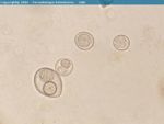
| + | [[Image:Isospora felis sporulated.jpg|thumb|right|150px|''Isospora felis'' sporulated - Courtesy of the Laboratory of Parasitology, University of Pennsylvania School of Veterinary Medicine]] | ||
| + | [[Image:Isospora felis unsporulated.jpg|thumb|right|150px|''Isospora felis'' unsporulated - Courtesy of the Laboratory of Parasitology, University of Pennsylvania School of Veterinary Medicine]] | ||
| + | *11 different Coccidia species although only two are of clinical significance | ||
| + | **Giant schizonts visible as white spots | ||
| + | |||
| + | *''Eimeria ovinoidalis'' | ||
| + | **Highly pathogenic | ||
| + | **[[Intestine Diarrhoea - Pathology|Diarrhoea]] | ||
| + | **Parasitises the [[Caecum - Anatomy & Physiology|caecum]] and [[Colon - Anatomy & Physiology|colon]] | ||
| + | |||
| + | *''Eimeria crandalis'' | ||
| + | **Varying pathogenicity | ||
| + | **Scours, grey, foul-smelling faeces | ||
| + | **Parasitises the [[Small Intestine - Anatomy & Physiology|small intestine]], [[Caecum - Anatomy & Physiology|caecum]] and [[Colon - Anatomy & Physiology|colon]] | ||
| + | |||
| + | *2 week prepatent period | ||
| + | |||
| + | *Disease frequently seen in lambs under 6 months old | ||
| + | **More often in twins and triplets when single lambs | ||
| + | |||
| + | *Oocyts from ewes (immune carriers) accumulate in poorly managed litter or around feed and water troughs | ||
| + | |||
| + | *Lambs born early in the year amplify the parasite problem increasing the parasite risk to lambs born later in the year | ||
| + | |||
| + | *Affected lambs may die before oocysts are found in the faeces | ||
| + | **Post-mortem diagnosis difficult | ||
| + | |||
| + | *Different species of ''Eimeria'' occurs in sheep and goats | ||
| + | |||
| + | *Infection may be coincident with ''Neospora'' or ''Cryptosporidium'' infections | ||
| + | **Mixed infections complicate the diagnosis as oocyst differentiation is difficult | ||
| + | |||
| + | *Other non-pathogenic species can cause papillomatous mucosal growths | ||
| + | |||
| + | '''Control''' | ||
| + | *Improve husbandry | ||
| + | **Avoid overcrowding | ||
| + | **Decrease stress | ||
| + | |||
| + | *Improve hygiene by dagging ewes | ||
| + | |||
| + | *Avoid mixing lambs of different ages | ||
| + | |||
| + | *Preventative measures include creep feeding lambs with decoquinate or oral dosing with diclazuril when lambs are 4-6 weeks | ||
| + | **A second dose can be given after 3 weeks | ||
| + | |||
| + | ==Coccidia of Goats== | ||
| + | [[Image:Goats.jpg|thumb|right|150px|Goats - nabrown RVC]] | ||
| + | [[Image:Eimeria leukarti horse.jpg|thumb|right|150px|''Eimeria leukarti'' - Joaquim Castellà Veterinary Parasitology Universitat Autònoma de Barcelona]] | ||
| + | [[Image:Isospora suis oocyst.jpg|thumb|right|150px|''Isospora suis'' oocyst from pig faeces - Joaquim Castellà Veterinary Parasitology Universitat Autònoma de Barcelona]] | ||
| + | [[Image:Isospora canis.jpg|thumb|right|150px|''Isospora canis'' - Joaquim Castellà Veterinary Parasitology Universitat Autònoma de Barcelona]] | ||
| + | [[Image:Coccidia.jpg|thumb|right|150px|Coccidia in Cat Faeces - Joel Mills]] | ||
| + | [[Image:Isospora felis.jpg|thumb|right|150px|''Isospora felis'' - Joaquim Castellà Veterinary Parasitology Universitat Autònoma de Barcelona]] | ||
| + | *Many ''Eimeria'' species | ||
| + | |||
| + | *2 ''Eimeria'' are pathogenic | ||
| + | **Cause [[Intestine Diarrhoea - Pathology|diarrhoea]] and a decreased growth rate | ||
| + | |||
| + | *Different species of ''Eimeria'' occurs in sheep and goats | ||
| + | |||
| + | ==Coccidia of Horses== | ||
| + | |||
| + | *Only one atypical ''Eimeria'' | ||
| + | |||
| + | *Forms large subepithelial gametocytes in villi | ||
| + | |||
| + | *Large, dark coloured oocysts | ||
| + | **Approximately 12μm | ||
| + | |||
| + | *Occasionally causes [[Intestine Diarrhoea - Pathology|diarrhoea]] | ||
| + | |||
| + | *''Besnoitia bennetti'' in [[Respiratory Parasitic Infections - Pathology#Besnoitia bennetti|larynx]] of horses | ||
| + | |||
| + | ==Coccidia of Pigs== | ||
| + | *Many species of ''Eimeria'' and ''Isospora'' | ||
| + | |||
| + | *Only ''Isospora suis'' is of clinical pathogenic importance | ||
| + | |||
| + | *Causes sporadic, serious and sometimes fatal disease in unweaned piglets | ||
| + | **Causes profuse [[Intestine Diarrhoea - Pathology|diarrhoea]] | ||
| + | |||
| + | *Very short 1 week prepatent period | ||
| + | |||
| + | *[[Intestine Diarrhoea - Pathology|Diarrhoea]] starts before oocysts are shed in faeces | ||
| + | **Ante-mortem diagnosis is difficult | ||
| + | |||
| + | *Death usually occurs after parasites have left the host | ||
| + | **Post-mortem diagnosis difficult | ||
| + | **''Isospora'' infections are '''self-limiting''' | ||
| + | |||
| + | ==Coccidia of Dogs== | ||
| + | *2 common and 2 less common ''Isospora'' species | ||
| + | |||
| + | *Occasionally can cause disease | ||
| + | |||
| + | *Little pathogenicity | ||
| + | |||
| + | *Even if faecal oocyst count is high, other causes of [[Intestine Diarrhoea - Pathology|diarrhoea]] should be looked for | ||
| + | |||
| + | *''Hepatozoon americanum'' and subclinical ''H. canis'' in [[Bones Hyperplastic and Neoplastic - Pathology#Hepatozoon|periosteal bone formation]] | ||
| + | **Both are Tick borne diseases | ||
| + | ***''H. canis'' – ''Rhipicephalus sanguineus'' | ||
| + | ***Ticks become infected by ingesting a blood meal containing macrophages and [[Neutrophils - WikiBlood|neutrophils]] infected with the parasite gamonts -> sexual replication in the gut of the tick -> oocysts containing infective sporozoites -> dogs ingest the tick schizogony occurs in numerous tissues | ||
| + | |||
| + | ==Coccidia of Cats== | ||
| + | *2 common ''Isospora'' species with little clinical significance | ||
| + | |||
| + | *Oocysts in faeces have to be distinguised from those of ''Toxoplasma'' (smaller) and ''Sarcocytis'' (sporulated or naked sporocyts in faeces) | ||
| + | |||
| + | ==Coccidia of Rabbits== | ||
| + | *3 pathogenic ''Eimeria'' species | ||
| + | **2 in the [[Caecum - Anatomy & Physiology|caecum]] | ||
| + | **1 in the bile duct | ||
| + | |||
| + | *''Eimeria steidae'' | ||
| + | **Parasitises the bile duct epithelium | ||
| + | **Travels via the bile duct to the [[Liver - Anatomy & Physiology|liver]] where it forms large white nodules | ||
| + | **Oocysts travel in the bile and are passed out in the faeces | ||
| + | **Causes ascites, [[Intestine Diarrhoea - Pathology|diarrhoea]], weight loss and polyuria | ||
| + | |||
| + | *Serious disease of both pet and farmed rabbits | ||
| + | |||
| + | *Treatment is by administration of drugs in drinking water | ||
| + | **E.g. Toltrazuril | ||
| + | |||
| + | *Hygiene is the best method of prevention to prevent sporocysts from sporulating | ||
| + | |||
| + | *Medicated feed can be used in commercial units | ||
| + | **E.g. Rabenidine | ||
| + | |||
| + | ==[[Protozoa Flashcards - Wikibugs#Coccidia|Coccidia Flashcards]]== | ||
| + | [[Category:Coccidia]] | ||
Revision as of 21:53, 8 April 2010
Introduction
- The oocyst is the resistant stage in the environment
- The infective sporozoite is released from the oocyst
- Inside the host, the sporozoites invade the intestinal epithelial tissue
- Sporozoites feed and grow
- As the sporozoite grows the nucleus divides forming a schizont
- The schizont contains numerous elongated merozoites
- The formation of merozoites is the first asexual reproductive stage called schizogony
- The schizont ruptures releasing the merozoites which also invade the epithelial cells
- Another generation of schizonts form which is the beginning of the sexual phase of reproduction called gametogony
- The merozoites form male microgamonts or female macrogamonts
- Collectively known as gamonts or gametocytes
- The microgamonts released from the microgametocyte penetrate and fertilise the macrogamont (which is contained within the macrogametocyte)
- Gametogony forms the zygote
- Surrounded by a cyst wall
- Forms the oocyst
- The oocyst is passed in the faeces and is unsporulated
- The oocyst becomes sporulated in the second asexual reproductive phase called sporogony
- Once the oocyst is sporulated it is infective
Coccidia of Cattle
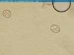
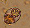
- Many species affect cattle
- Cattle under a year old are usually infected sporadically
- 2-3 week prepatent period
- Eimeria bovis
- Eimeria zuernii
- Endogenous stages in connective tissue of lamina propria of the lower small intestine and in the epithelial cells of the caecum and colon
- More pathogenic than Eimeria bovis
- Causes blood stained dysentery, tenesmus and sloughed mucosa
- Oocysts are spherical and measure 16μm
- Mainly occurs in calves in poor conditions and bought-in calves
- Also occurs in suckler calves turned out in spring
- Eimeria alabamensis associated with diarrhoea in calves after spring turnout
- Passive immunity is sufficient during the neonatal period
- Can be concurrent with cryptosporidium, viral and bacterial agents
Diagnosis
- History, clinical signs, diarrhoea (often with blood) and a decrease in weight gain
- Post-mortem
- High faecal oocyst count
- However, healthy animals can pass millions of oocysts from mixed species infections which have no pathogenic significance
- Animals may die before oocysts are shed
Control
- Improve husbandry
- Improve sanitation
- Increase bedding
- Raise food and water troughs to avoid faecal contamination
- Preventative in-feed medication
- E.g. Decoquinate
- Injectable antiprotozoals may limit oocyst production but animals should still be moved to a clean environment
- E.g. Sulphamethoxypyridazine
Coccidia of Sheep
- 11 different Coccidia species although only two are of clinical significance
- Giant schizonts visible as white spots
- Eimeria crandalis
- Varying pathogenicity
- Scours, grey, foul-smelling faeces
- Parasitises the small intestine, caecum and colon
- 2 week prepatent period
- Disease frequently seen in lambs under 6 months old
- More often in twins and triplets when single lambs
- Oocyts from ewes (immune carriers) accumulate in poorly managed litter or around feed and water troughs
- Lambs born early in the year amplify the parasite problem increasing the parasite risk to lambs born later in the year
- Affected lambs may die before oocysts are found in the faeces
- Post-mortem diagnosis difficult
- Different species of Eimeria occurs in sheep and goats
- Infection may be coincident with Neospora or Cryptosporidium infections
- Mixed infections complicate the diagnosis as oocyst differentiation is difficult
- Other non-pathogenic species can cause papillomatous mucosal growths
Control
- Improve husbandry
- Avoid overcrowding
- Decrease stress
- Improve hygiene by dagging ewes
- Avoid mixing lambs of different ages
- Preventative measures include creep feeding lambs with decoquinate or oral dosing with diclazuril when lambs are 4-6 weeks
- A second dose can be given after 3 weeks
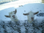
Coccidia of Goats
- Many Eimeria species
- 2 Eimeria are pathogenic
- Cause diarrhoea and a decreased growth rate
- Different species of Eimeria occurs in sheep and goats
Coccidia of Horses
- Only one atypical Eimeria
- Forms large subepithelial gametocytes in villi
- Large, dark coloured oocysts
- Approximately 12μm
- Occasionally causes diarrhoea
- Besnoitia bennetti in larynx of horses
Coccidia of Pigs
- Many species of Eimeria and Isospora
- Only Isospora suis is of clinical pathogenic importance
- Causes sporadic, serious and sometimes fatal disease in unweaned piglets
- Causes profuse diarrhoea
- Very short 1 week prepatent period
- Diarrhoea starts before oocysts are shed in faeces
- Ante-mortem diagnosis is difficult
- Death usually occurs after parasites have left the host
- Post-mortem diagnosis difficult
- Isospora infections are self-limiting
Coccidia of Dogs
- 2 common and 2 less common Isospora species
- Occasionally can cause disease
- Little pathogenicity
- Even if faecal oocyst count is high, other causes of diarrhoea should be looked for
- Hepatozoon americanum and subclinical H. canis in periosteal bone formation
- Both are Tick borne diseases
- H. canis – Rhipicephalus sanguineus
- Ticks become infected by ingesting a blood meal containing macrophages and neutrophils infected with the parasite gamonts -> sexual replication in the gut of the tick -> oocysts containing infective sporozoites -> dogs ingest the tick schizogony occurs in numerous tissues
- Both are Tick borne diseases
Coccidia of Cats
- 2 common Isospora species with little clinical significance
- Oocysts in faeces have to be distinguised from those of Toxoplasma (smaller) and Sarcocytis (sporulated or naked sporocyts in faeces)
Coccidia of Rabbits
- 3 pathogenic Eimeria species
- 2 in the caecum
- 1 in the bile duct
- Eimeria steidae
- Serious disease of both pet and farmed rabbits
- Treatment is by administration of drugs in drinking water
- E.g. Toltrazuril
- Hygiene is the best method of prevention to prevent sporocysts from sporulating
- Medicated feed can be used in commercial units
- E.g. Rabenidine

