Difference between revisions of "Fertilisation - Anatomy & Physiology"
Jump to navigation
Jump to search
| (5 intermediate revisions by the same user not shown) | |||
| Line 1: | Line 1: | ||
| + | {{toplink | ||
| + | |backcolour =EED2EE | ||
| + | |linkpage =Reproductive System - Anatomy & Physiology | ||
| + | |linktext =Reproductive System | ||
| + | |maplink = Reproductive System (Content Map) - Anatomy & Physiology | ||
| + | |pagetype =Anatomy | ||
| + | |sublink1=Reproductive System - Anatomy & Physiology#Fertilisation.2C Implantation and Early Embryonic Development | ||
| + | |subtext1=FERTILISATION , IMPLANTATION AND EARLY EMBRYONIC DEVELOPMENT''' | ||
| + | }} | ||
| + | <br> | ||
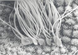
[[Image:Fertilization histology.jpg|right|thumb|250px|<small><center> Electron Scanning Micrograph of Spermatozoa in the Uterine Horn. Copyright RVC 2008 (Courtesy of John Bredl (RVC))</center></small>]] | [[Image:Fertilization histology.jpg|right|thumb|250px|<small><center> Electron Scanning Micrograph of Spermatozoa in the Uterine Horn. Copyright RVC 2008 (Courtesy of John Bredl (RVC))</center></small>]] | ||
| Line 4: | Line 14: | ||
* When the spermatozoon completely penetrates the zona pellucida and reaches the perivitelline space, it settles into a bed of '''microvilli''' formed by the oocyte plasma membrane. | * When the spermatozoon completely penetrates the zona pellucida and reaches the perivitelline space, it settles into a bed of '''microvilli''' formed by the oocyte plasma membrane. | ||
* Oocyte plasma membrane fuses with the '''equitorial segment''' and the fertilizing spermatozoon is engulfed. | * Oocyte plasma membrane fuses with the '''equitorial segment''' and the fertilizing spermatozoon is engulfed. | ||
| − | **Brought about by a fusion protein that is inactive prior to the [[ | + | **Brought about by a fusion protein that is inactive prior to the [[Copulation_-Sperm_in_the_Female_Tract_-_Anatomy_%26_Physiology|acrosome reaction]]. |
* Nucleus of the spermatozoon is within the oocyte cytoplasm. | * Nucleus of the spermatozoon is within the oocyte cytoplasm. | ||
* Sperm nuclear membrane disappears. | * Sperm nuclear membrane disappears. | ||
| Line 51: | Line 61: | ||
''N.B: Although sperm mitochondria enters the Ooctye cytoplasm, they carry a ubiquitin signal. They are subsequently destroyed by Ubiquiitinase and do NOT contribute to mitochondrial DNA. Therefore mitochondial DNA is entirely from the maternal side.'' | ''N.B: Although sperm mitochondria enters the Ooctye cytoplasm, they carry a ubiquitin signal. They are subsequently destroyed by Ubiquiitinase and do NOT contribute to mitochondrial DNA. Therefore mitochondial DNA is entirely from the maternal side.'' | ||
| − | |||
| − | |||
| − | |||
| − | |||
| − | |||
Revision as of 18:01, 16 June 2010
|
|
Fusion with the Oocyte
- When the spermatozoon completely penetrates the zona pellucida and reaches the perivitelline space, it settles into a bed of microvilli formed by the oocyte plasma membrane.
- Oocyte plasma membrane fuses with the equitorial segment and the fertilizing spermatozoon is engulfed.
- Brought about by a fusion protein that is inactive prior to the acrosome reaction.
- Nucleus of the spermatozoon is within the oocyte cytoplasm.
- Sperm nuclear membrane disappears.
- Sperm nucleus decondenses.
Cortical Reaction - Block to Polyspermy
- During the first and second meiotic divisions of oogenesis small, dense cortical granules move to the periphery of the Oocyte cytoplasm.
- Cortical granules consist of:
- Mucopolysaccharides
- Proteases
- Peroxidase
- Cortical granules undergo exocytosis, releasing their contents into the perivitteline space.
- Contents of cortical granules cross-links zona proteins to make them impenetrable to further spermatozoa.
- This is known as the zona block, it prevents polyspermy (fertilization by more than one sperm)which would result in embryo death.
- The cortical reaction also reduces the ability of the oocyte plasma membrane to fuse with additional spermatozoa.
- This is the vitelline block to polyspermy.
- If two spermatozoa enter the perivitelline space simultaneously, they both contact the oocyte and proteins are not cross-linked rapidly enough to block penetration of the zona pellucida.
- Thus, the block depends on limiting the number of Spermatozoa in the vicinity of the Oocyte.
- Only a small sub-population are released from the store in the female tract over the period of ovulation so that 7-10 out of the original millions are around the oocyte at the period of fertilization.
Formation of Pronuclei
After the sperm nucleus enters the Oocyte it becomes the male pronucleus. Before this can occur, the sperm nucleus must undergo marked changes within the Oocyte cytoplasm.
- Disulphide cross-links render the nucleus almost inert. This is important when exposed to the hostile environment of the female tract during transport and also on penetration through the Zona Pellucida.
- In the Oocyte cytoplasm, the nucleus must decondense in order for male and female chromosomes to pair.
- Disulphide cross-links are rapidly reduced in the Oocyte cytoplasm primarily by Glutathione.
Syngamy
- Male and female pronuclei fuse.
- Sperm delivers a haploid complement of chromosomes, but also activates the Oocyte by:
- A unique phospholipase Cζ (zeta) causes release of calcium from the endoplasmic reticulem to the cytosol. This is then re-sequestered and bound in the endoplasmic reticulem to give regular 'spikes' or oscillations of calcium that signal rapidly across the Oocyte. The calcium signal activates the Oocyte from meiotic arrest.
- Provides the centriole contribution to initial cell division, allowing formation of the two-celled embryo.
N.B: Although sperm mitochondria enters the Ooctye cytoplasm, they carry a ubiquitin signal. They are subsequently destroyed by Ubiquiitinase and do NOT contribute to mitochondrial DNA. Therefore mitochondial DNA is entirely from the maternal side.
