Difference between revisions of "Fasciola hepatica"
| Line 9: | Line 9: | ||
''Fasciola Hepatica'' is an hepatic parasite found in mainly in ruminants, namely cows, sheep and goats, but also known to affect horses and pigs. It is found Worldwide, and within the UK, with its prevalence ever increasing. It is responsible for a 10-15% production loss in each infected animal, as it affects meat, milk and wool production, so is of huge economic consequence. | ''Fasciola Hepatica'' is an hepatic parasite found in mainly in ruminants, namely cows, sheep and goats, but also known to affect horses and pigs. It is found Worldwide, and within the UK, with its prevalence ever increasing. It is responsible for a 10-15% production loss in each infected animal, as it affects meat, milk and wool production, so is of huge economic consequence. | ||
| − | ''Fasiola Hepatica'' has a definitive ruminant mammalian host and an intermediate | + | ''Fasiola Hepatica'' has a definitive ruminant mammalian host and an intermediate molluscan host. Within Europe the intermediate host is almost exclusively the snail 'Lymnaea truncatulata'. The snail habitat is crucial to the survival of the parasite, so wet conditions are favourable to the development and spread of Fasciola hepatica |
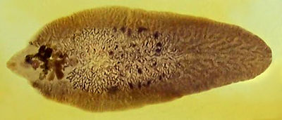
[[Image:Fasciola hepatica.jpg|400px|thumb|right|'''Fasciola hepatica (Copyright Adam Cuerden, Wikimedia Commons) ''']] | [[Image:Fasciola hepatica.jpg|400px|thumb|right|'''Fasciola hepatica (Copyright Adam Cuerden, Wikimedia Commons) ''']] | ||
| Line 46: | Line 46: | ||
===Pathogenesis=== | ===Pathogenesis=== | ||
| − | The severity of the infection is mainly dependent on the number of metacercariae ingested. The Pathogenesis is often described as two-fold. The first stage | + | The severity of the infection is mainly dependent on the number of metacercariae ingested. The Pathogenesis is often described as two-fold. The first stage occurring when the parasite migrates through the liver parenchyma, causing liver damage and haemorrhage. The second phase occurs when the parasite is in the bile ducts, and damage is a result of the haematophagic activity of the adult flukes. |
===Hosts=== | ===Hosts=== | ||
| Line 58: | Line 58: | ||
===Life Cycle=== | ===Life Cycle=== | ||
| − | Adult flukes in the bile ducts shed eggs directly into the | + | Adult flukes in the bile ducts shed eggs directly into the bile, which then subsequently enter the intestine. Eggs are then passes out in the faeces of the mammalian host, where they develop and hatch releasing motile ciliated miracidia. These require 9-10 days at optimal temperatures, of around 22-26 degrees. The miracidium have a short life and must locate a suitable snail, the intermediate host, within approximately 3 hours if they are to be effective and continue the life cycle. |
| − | If | + | If successful, the miracidium will then develop into sporocysts, then enter the redial stages to the final stages within the intermediate host, which is development into cercaria. These cercaria are then released from the snail, and attach to surfaces such as the tips of grass. Here they encyst and form metacercaria. This represents the infective stage of the lifecycle. |
Development from miracidium into metacercariae takes around 6-7 weeks under favourable conditions, however, this period can be much longer in unfavourable conditions. | Development from miracidium into metacercariae takes around 6-7 weeks under favourable conditions, however, this period can be much longer in unfavourable conditions. | ||
| Line 87: | Line 87: | ||
In temperate areas, there are two superimposed epidemiological cycles, known as the summer and winter infections of the snail. On mainland Britain, the summer cycle predominates as a high proportion of snails perish during the winter, but very occasionally, weather sequences allow the winter cycle to affect the pattern of disease. On the west coast of Ireland, the winter cycle of events determines the timing of clinical outbreaks. | In temperate areas, there are two superimposed epidemiological cycles, known as the summer and winter infections of the snail. On mainland Britain, the summer cycle predominates as a high proportion of snails perish during the winter, but very occasionally, weather sequences allow the winter cycle to affect the pattern of disease. On the west coast of Ireland, the winter cycle of events determines the timing of clinical outbreaks. | ||
| − | === Summer infection of the snail === | + | ==== Summer infection of the snail ==== |
Fluke eggs passed in '''spring''' | Fluke eggs passed in '''spring''' | ||
| Line 102: | Line 102: | ||
→ immature flukes migrate through liver | → immature flukes migrate through liver | ||
| − | → '''acute''' disease '''September-November'''; or '''chronic''' | + | → '''acute''' disease '''September-November'''; or '''chronic''' disease '''January''' onwards |
| − | === Winter infection of the snail === | + | ==== Winter infection of the snail ==== |
Fluke eggs passed in '''late summer''' | Fluke eggs passed in '''late summer''' | ||
Revision as of 15:49, 10 July 2010
| Also known as: | Liver Fluke |
Introduction
Fasciola Hepatica is an hepatic parasite found in mainly in ruminants, namely cows, sheep and goats, but also known to affect horses and pigs. It is found Worldwide, and within the UK, with its prevalence ever increasing. It is responsible for a 10-15% production loss in each infected animal, as it affects meat, milk and wool production, so is of huge economic consequence.
Fasiola Hepatica has a definitive ruminant mammalian host and an intermediate molluscan host. Within Europe the intermediate host is almost exclusively the snail 'Lymnaea truncatulata'. The snail habitat is crucial to the survival of the parasite, so wet conditions are favourable to the development and spread of Fasciola hepatica
Scientific Classification
| Kingdom | Animalia |
| Phylum | Platyhelminthes |
| Class | Trematoda |
| Subclass | Digenea |
| Order | Echinostomida |
| Family | Fasciolidae |
| Genus | Fasciola |
| Species | F. Hepatica |
Pathogenesis
The severity of the infection is mainly dependent on the number of metacercariae ingested. The Pathogenesis is often described as two-fold. The first stage occurring when the parasite migrates through the liver parenchyma, causing liver damage and haemorrhage. The second phase occurs when the parasite is in the bile ducts, and damage is a result of the haematophagic activity of the adult flukes.
Hosts
Fasciola hepatica has an indirect life cycle, meaning it has both intermediate and final hosts.
Fasciola hepatica is seen most commonly in sheep, cattle, and goats, but may also be seen in horse, deer, and man.
The most important intermediate host within Europe is the snail of the genus Lymnaea. The most common, Lymnaea truncatula, which is an amphibious snail found worldwide.
Life Cycle
Adult flukes in the bile ducts shed eggs directly into the bile, which then subsequently enter the intestine. Eggs are then passes out in the faeces of the mammalian host, where they develop and hatch releasing motile ciliated miracidia. These require 9-10 days at optimal temperatures, of around 22-26 degrees. The miracidium have a short life and must locate a suitable snail, the intermediate host, within approximately 3 hours if they are to be effective and continue the life cycle.
If successful, the miracidium will then develop into sporocysts, then enter the redial stages to the final stages within the intermediate host, which is development into cercaria. These cercaria are then released from the snail, and attach to surfaces such as the tips of grass. Here they encyst and form metacercaria. This represents the infective stage of the lifecycle.
Development from miracidium into metacercariae takes around 6-7 weeks under favourable conditions, however, this period can be much longer in unfavourable conditions.
The final host, or the definitive host then ingests the metacercariae from the grass on the pasture, and these pass through the body into the intestine, where they excyst in the wall of the small intestine. They then travel through the wall of the gut, and migrate into the liver, through the liver parenchyma. The young liver flukes migrate through the liver for around 6-8 weeks before entering the bile ducts. They may also migrate into the gall bladder, where they reach full sexual maturity.
The prepatent period of Fasciola hepatica is 10-12 weeks. In untreated sheep it may survive and continue to infect for many years. In cattle it is usually less than 1 year.
Snail biology
Lymnaea truncatula
- 5-10mm long
- Brown-black shell with 5-6 spirals
- 1st spiral is greater than half the total length
- The shell opens on the right (when held with the opening upwards)
- Feeds on green slime
- Multiplies rapidly when food is abundant
- Most die during the British winter (unless very mild)
- Survivors lay eggs in spring, which hatch in June
Habitats
- Lymnaea are found in muddy areas (but not on highly acidic soils)
- Habitats may be permanent (dry summer) or temporary (wet summer)
Epidemiology
In temperate areas, there are two superimposed epidemiological cycles, known as the summer and winter infections of the snail. On mainland Britain, the summer cycle predominates as a high proportion of snails perish during the winter, but very occasionally, weather sequences allow the winter cycle to affect the pattern of disease. On the west coast of Ireland, the winter cycle of events determines the timing of clinical outbreaks.
Summer infection of the snail
Fluke eggs passed in spring
→ hatch in June (i.e. coincident with snail hatch)
→ miracidia infect newly hatched snails
→ develop and multiply in snail hepatopancreas during summer
→ cercariae shed from late August onwards
→ metacercariae ingested by sheep
→ immature flukes migrate through liver
→ acute disease September-November; or chronic disease January onwards
Winter infection of the snail
Fluke eggs passed in late summer
→ infect snails
→ development halted when temperature <10°C (i.e. flukes trapped in hibernating snails through the winter)
→ development resumes when temperature >10°C
→ cercariae shed from July
→ disease from August
References
Taylor, M.A, Coop, R.L., Wall,R.L. (2007) Veterinary Parasitology Blackwell Publishing
G.L. Pritchard et al., Emergence of fasciolosis in cattle in East Anglia, The Veterinary Record, November 5, 2005.

