Difference between revisions of "Sarcocystis"
Jump to navigation
Jump to search
| (2 intermediate revisions by 2 users not shown) | |||
| Line 59: | Line 59: | ||
*Cause meat inspection losses | *Cause meat inspection losses | ||
| − | *''Sarcocystis'' in [[ | + | *''Sarcocystis'' in [[Muscles Inflammatory - Pathology#Protozoa|myositis]] |
*Experimental infections cause severe, acute pyrexic disease when the organism multiplies in the vascular endothelium | *Experimental infections cause severe, acute pyrexic disease when the organism multiplies in the vascular endothelium | ||
| Line 71: | Line 71: | ||
[[Image:Sarcocyst in muscle.jpg|right|thumb|100px|<small><center>Sarcocyst (Image sourced from Bristol Biomed Image Archive with permission)</center></small>]] | [[Image:Sarcocyst in muscle.jpg|right|thumb|100px|<small><center>Sarcocyst (Image sourced from Bristol Biomed Image Archive with permission)</center></small>]] | ||
**Histologically: | **Histologically: | ||
| − | ***Merozoites causing focal and segmental [[ | + | ***Merozoites causing focal and segmental [[Muscles Degenerative - Pathology#Necrosis|necrosis]] |
| − | ***May involve [[ | + | ***May involve [[Muscles Degenerative - Pathology#Calcification|mineralisation]] |
***Non-purulent myositis with plasma cells, lymphocytes and macrophages | ***Non-purulent myositis with plasma cells, lymphocytes and macrophages | ||
**Diaphragm and masseters most severely affected | **Diaphragm and masseters most severely affected | ||
| − | |||
| − | |||
| − | |||
| − | |||
| − | |||
[[Category:Tissue_Cyst_Forming_Coccidia]] | [[Category:Tissue_Cyst_Forming_Coccidia]] | ||
[[Category:To_Do_-_Parasites]] | [[Category:To_Do_-_Parasites]] | ||
Revision as of 10:12, 22 July 2010
- Most infections are asymptomatic
- Heavy infections are causes of chronic wasting in large animals, hide condemnation and downgrading of carcasses
- Sarcocystis should be differentiated from other tissue-cyst forming coccidia
- There are many species of Sarcocystis which differ in size from microscopic to several centimetres in length
- S.neurona is an important equine pathogen in the USA
- Infective cyst in the intermediate host is called a sarcocyst
Life Cycle
- The individual life cycle of some species is incompletely understood
- Indirect life cycle
- Life cycle alternates between the final and the obligatory intermediate host
- Only one final and one intermediate host
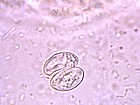
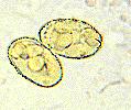
- Sporulated oocyst has 2 sporocysts containing 4 sporozoites
- Naked sporocyst usually seen in faeces as the oocyst wall is very delicate
- Oocyst measures 15μm in length
- No schizogony in final host
- Gametogeny occurs deep in subepithelial tissue
- Faecal oocyst count is low
- Oocysts are sporulated when passed
- Difficult to find on faecal examination as the sporocysts are few in number and small
- Ingestion of sporocyst by intermediate host
- 2 phases of rapid asexual reproduction in vascular endothelial cells
- Slow multiplication of bradyzoites in muscle tissue
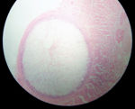
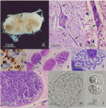
- Sarcocyst forms with bradyzoites inside, surrounded by a cyst wall and divided into compartments
Epidemiology
- Final hosts are carnivores and omnivores
- Intermediate hosts are herbivores and omnivores
- Humans are the final host for some species and the intermediate hosts for others
- Final host for species infecting cattle and pigs
- Dogs are final hosts for species infecting cattle, sheep, goats, pigs and horses
- Cats are final hosts for species infecting cattle, sheep and pigs
Pathogenesis
- Widespread infection but mostly asymptomatic
- Cause meat inspection losses
- Sarcocystis in myositis
- Experimental infections cause severe, acute pyrexic disease when the organism multiplies in the vascular endothelium
- Can cause chronic wasting disease in cattle and horses
- Causes abortion and post-natal disease in sheep
- Myositis
- Histologically:
- Merozoites causing focal and segmental necrosis
- May involve mineralisation
- Non-purulent myositis with plasma cells, lymphocytes and macrophages
- Diaphragm and masseters most severely affected
- Histologically:


