Difference between revisions of "Small Animal Emergency and Critical Care Medicine: Self-Assessment Color Review, Second Edition, Q&A 16"
(Created page with "{{CRC Press}} {{Student tip |X = an example using clear imagery with interactive use of arrows. }} <br><br><br> File:Small Animal Emergency and Critical Care Medicine 2E Q16...") |
|||
| (One intermediate revision by the same user not shown) | |||
| Line 28: | Line 28: | ||
[[Category:CRC Press flashcards]] | [[Category:CRC Press flashcards]] | ||
| − | To purchase the full text with your 20% | + | To purchase the full text with your 20% discount, go to the [https://www.crcpress.com/9781482225921 CRC Press] Veterinary website and use code VET18. |
| + | <br> <br> | ||
{{#tag:imagemap|Image:Next Question CRC Press.png{{!}}center{{!}}200px | {{#tag:imagemap|Image:Next Question CRC Press.png{{!}}center{{!}}200px | ||
| − | rect 0 0 860 850 [[Small Animal Emergency and Critical Care Medicine: Self-Assessment Color Review, Second Edition, | + | rect 0 0 860 850 [[Small Animal Emergency and Critical Care Medicine: Self-Assessment Color Review, Second Edition, Q&A 17|Next question]] |
desc none}} | desc none}} | ||
Latest revision as of 09:38, 26 November 2018
| This question was provided by CRC Press. See more case-based flashcards |

|
Student tip: This case is an example using clear imagery with interactive use of arrows. |
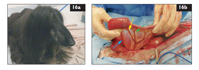
A 3-year-old male neutered Afghan hound presents for persistent vomiting over the past 2 days, not eating, and looking at his abdomen (16a). He has a history of eating things out of the trash and then developing diarrhea, but he has never been hospitalized. T = 39.2°C (102.6°F); HR = 165 bpm; RR = 30 bpm; CRT = 3 sec; MM pale pink, dry; femoral pulses weak. Abdominal palpation demonstrates a firm 3 cm × 2 cm mass within the small intestines in the mid-abdominal region. Thoracic auscultation findings normal. Radiographs show evidence of an intestinal obstruction. The dog is volume replaced and prepared for anesthesia.
| Question | Answer | Article | |
| During surgery, the mid jejunum containing a FB is exteriorized and an intestinal resection–anastomosis performed without an enterotomy. Which arrow points to the vessel that will be ligated during the resection (16b)? | The green one.
|
Link to Article | |
| Which intestinal anastomosis suturing technique (interrupted or continuous) provides better appositional closure and less leakage? | Simple continuous is better. Anastomotic leakage is reported in 3% of animals undergoing continuous sutured anastomosis and up to 11% of animals undergoing interrupted sutured anastomosis.
|
Link to Article | |
| What suture pattern can reduce eversion of mucosal tissue during intestinal anastamosis? | A modified Gambee suture pattern: the needle is passed full thickness through the intestinal wall and then back through the mucosa on the near side. Then the needle is placed into the mucosa–submucosa border on the far side, pushing the mucosa into the lumen, and then passed full thickness back out that side.
|
Link to Article | |
| How can the anastomosis site be checked for leaks? | Distend the segment with sterile saline injected into the lumen while continuing to occlude the intestinal segments on both sides distal to the anastomotic site.
|
Link to Article | |
| What are the risk factors associated with the complication of leakage following anastamosis? | FB removal or resection of traumatized intestines, pre-existing peritonitis, hypoalbuminemia, reduced blood supply.
|
Link to Article | |
To purchase the full text with your 20% discount, go to the CRC Press Veterinary website and use code VET18.
