Difference between revisions of "Cytology Q&A 16"
Jump to navigation
Jump to search
Ggaitskell (talk | contribs) |
|||
| (2 intermediate revisions by 2 users not shown) | |||
| Line 1: | Line 1: | ||
| − | {{Template:Manson}} | + | {{Template:Manson |
| + | |book = Cytology Q&A}} | ||
| − | [[Image:|centre|500px]] | + | [[Image:Cytology 16.jpg|centre|500px]] |
<br /> | <br /> | ||
| Line 16: | Line 17: | ||
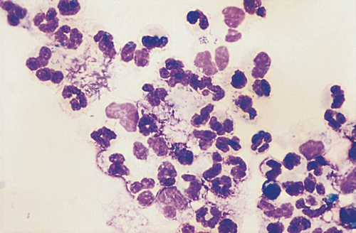
*However, the outline of neutrophils is visible in some cases and the disrupted cells are considered to be karyolytic neutrophils. | *However, the outline of neutrophils is visible in some cases and the disrupted cells are considered to be karyolytic neutrophils. | ||
*No microorganisms are seen. | *No microorganisms are seen. | ||
| − | |l1= | + | |l1=Exudate#Cytology |
|q2=What is the fluid classification? | |q2=What is the fluid classification? | ||
|a2= | |a2= | ||
An exudate. | An exudate. | ||
| − | |l2= | + | |l2=Exudate#Cytology |
|q3=What is your diagnosis? | |q3=What is your diagnosis? | ||
|a3=Acute peritonitis. <br><br> | |a3=Acute peritonitis. <br><br> | ||
Given the history and clinical signs, this is suggestive of foreign body reticuloperitonitis. <br><br> | Given the history and clinical signs, this is suggestive of foreign body reticuloperitonitis. <br><br> | ||
Note: Reference values for NCCs and classification of effusions in large animals differ from those for small animal specimens. | Note: Reference values for NCCs and classification of effusions in large animals differ from those for small animal specimens. | ||
| − | |l3= | + | |l3=Traumatic Pericarditis |
</FlashCard> | </FlashCard> | ||
Latest revision as of 16:01, 23 September 2011
| This question was provided by Manson Publishing as part of the OVAL Project. See more Cytology Q&A. |
Three days after calving, a four-year-old Friesian cow became acutely ill, with brisket and submandibular oedema. She was stiff and grunted with respiration. Peritoneal fluid withdrawn from a site 5 cm cranial and 5 cm medial to the milk well had a TP of 48 g/l and an NCC of 37 ×109/l. A cytospun smear of the peritoneal fluid was prepared (Wright–Giemsa, ×50 oil).
| Question | Answer | Article | |
| What cells are present? |
|
Link to Article | |
| What is the fluid classification? | An exudate. |
Link to Article | |
| What is your diagnosis? | Acute peritonitis. Given the history and clinical signs, this is suggestive of foreign body reticuloperitonitis. |
Link to Article | |
