Difference between revisions of "Feline Medicine Q&A 02"
Ggaitskell (talk | contribs) (Created page with "{{Template:Manson Sparkes}} [[Image:|centre|500px]] <br /> '''A 6-year-old neutered male DSH cat presents with crusting lesions around the mouth (184), ears, ventral abdomen, ...") |
|||
| (4 intermediate revisions by 3 users not shown) | |||
| Line 1: | Line 1: | ||
{{Template:Manson Sparkes}} | {{Template:Manson Sparkes}} | ||
| − | [[Image:|centre|500px]] | + | [[Image:Feline Medicine 02.png|centre|500px]] |
<br /> | <br /> | ||
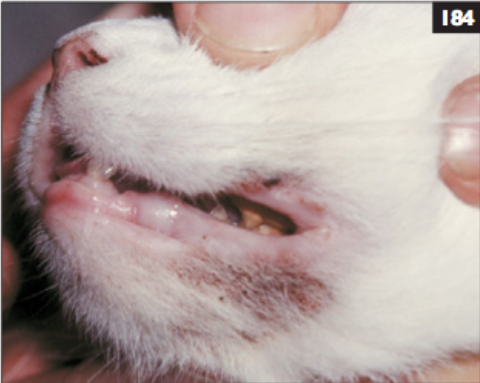
| − | '''A 6-year-old neutered male DSH cat presents with crusting lesions around the mouth | + | '''A 6-year-old neutered male DSH cat presents with crusting lesions around the mouth, ears, ventral abdomen, and nail beds. Histopathology reveals subcorneal pustules with acantholytic keratinocytes.''' |
<br /> | <br /> | ||
| Line 13: | Line 13: | ||
|a1= | |a1= | ||
The clinical signs and histopathology in this cat are typical of pemphigus foliaceous. | The clinical signs and histopathology in this cat are typical of pemphigus foliaceous. | ||
| − | |l1= | + | |l1=Pemphigus Foliaceus |
|q2=What is its cause and how should it be treated? | |q2=What is its cause and how should it be treated? | ||
|a2= | |a2= | ||
| Line 24: | Line 24: | ||
However, these are relatively superficial in the epidermis and are very fragile, so are rarely seen. <br><br> | However, these are relatively superficial in the epidermis and are very fragile, so are rarely seen. <br><br> | ||
Erosions and ulcers with crusting and exudation are therefore the common signs. Cytology of exudate may be helpful diagnostically as it may reveal the rounded acantholytic keratinocytes typical of the disease, and immunofluorescence can be used to demonstrate the deposition of antibodies in the lesions. | Erosions and ulcers with crusting and exudation are therefore the common signs. Cytology of exudate may be helpful diagnostically as it may reveal the rounded acantholytic keratinocytes typical of the disease, and immunofluorescence can be used to demonstrate the deposition of antibodies in the lesions. | ||
| − | |l2= | + | |l2=Pemphigus Foliaceus |
|q3=What is the prognosis for the cat? | |q3=What is the prognosis for the cat? | ||
|a3= | |a3= | ||
| Line 30: | Line 30: | ||
Glucocorticoids are the treatment of choice (e.g. 2–4 mg/kg/day oral prednisolone, followed by a reducing dose when in remission). <br><br> | Glucocorticoids are the treatment of choice (e.g. 2–4 mg/kg/day oral prednisolone, followed by a reducing dose when in remission). <br><br> | ||
If glucocorticoid-sparing therapy is needed, chlorambucil often produces good results. | If glucocorticoid-sparing therapy is needed, chlorambucil often produces good results. | ||
| − | |l3= | + | |l3=Pemphigus Foliaceus#Treatment |
</FlashCard> | </FlashCard> | ||
Latest revision as of 15:58, 30 September 2011
| This question was provided by Manson Publishing as part of the OVAL Project. See more Feline Medicine questions |
A 6-year-old neutered male DSH cat presents with crusting lesions around the mouth, ears, ventral abdomen, and nail beds. Histopathology reveals subcorneal pustules with acantholytic keratinocytes.
| Question | Answer | Article | |
| What is this disease? | The clinical signs and histopathology in this cat are typical of pemphigus foliaceous. |
Link to Article | |
| What is its cause and how should it be treated? | Pemphigus foliaceous is an autoimmune skin disease with deposition of autoantibodies in the epidermis targeted against intercellular adhesion molecules (cadherin desmosomal glycoproteins) causing loss of cellular adhesion.
However, these are relatively superficial in the epidermis and are very fragile, so are rarely seen. |
Link to Article | |
| What is the prognosis for the cat? | The prognosis for pemphigus foliaceous is good with most cases responding well to immunosuppressive therapy, although prolonged and sometimes life-long therapy may be required. |
Link to Article | |
