Difference between revisions of "Oesophagus - Anatomy & Physiology"
| (41 intermediate revisions by 12 users not shown) | |||
| Line 1: | Line 1: | ||
| − | + | {{OpenPagesTop}} | |
| − | |||
==Introduction== | ==Introduction== | ||
| − | The oesophagus (or gullet) is a muscular tube which transports food from the pharynx to the stomach. A bolus of food is passed down the oesophagus by | + | The oesophagus (or gullet) is a muscular tube which transports food from the [[Pharynx - Anatomy & Physiology|pharynx]] to the [[Monogastric Stomach - Anatomy & Physiology|stomach]]. A bolus of food is passed down the oesophagus by peristalsis. The oesophagus is divided into cervical, thoracic and abdominal sections. |
| − | |||
| − | The oesophagus is | ||
| − | |||
==Structure and Function== | ==Structure and Function== | ||
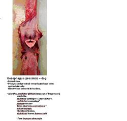
| + | [[Image:Oesophagus anatomy.jpg|thumb|right|250px|Oesophagus Anatomy - Copyright RVC 2008]] | ||
| + | The oesophagus begins dorsal to the '''cricoid cartilage''' of the [[Larynx - Anatomy & Physiology|larynx]]. It follows the trachea down the neck, first on the left and then medially once in thorax in the mediastinum. It passes over the [[Heart - Anatomy & Physiology|heart]] then through the oesophageal hiatus of the diaphragm. It passes over the dorsal border of the [[Liver - Anatomy & Physiology|liver]] then joins the [[Monogastric Stomach - Anatomy & Physiology|stomach]] at the cardia. The cervical section is accompanied by the common carotid artery, the vagosympathetic trunk and the recurrent laryngeal nerves. The thoracic section is accompanied by the right and left vagus nerves ([[Cranial Nerves - Anatomy & Physiology|CN X]]). | ||
| − | + | There are different proportions of striated muscle across the species; | |
| − | + | :Dog and ruminant = 100% | |
| − | + | :Cat = 80% (rostral) | |
| − | + | :Horse = 65% (rostral) | |
| − | + | :Pig = 33% (rostral) | |
| − | |||
| − | |||
| − | |||
| − | |||
| − | |||
| − | |||
| − | |||
| − | |||
| − | |||
| − | |||
| − | |||
| − | |||
| − | |||
| − | |||
| − | |||
==Histology== | ==Histology== | ||
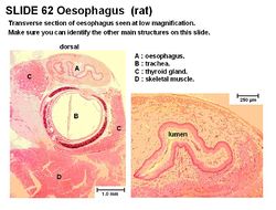
| + | [[Image:Oesophagus Histology.jpg|thumb|right|250px|Oesophagus Histology (Rat) - Copyright RVC 2008]] | ||
| + | The oesophagus has a '''stratified squamous epithelium'''. It has mucosal folds present for distension. The degree of keratinisation of the oesophagus depends on the animal's diet. | ||
| − | + | The '''lamina propria''' of the oesophagus contains collagen and elastic fibres that are sparsely distributed. The '''lamina muscularis''' of the oesophagus is smooth or skeletal muscle, depending on the species. The inner circular layer of the '''tunica muscularis''' thickens near the gastric junction, forming a sphincter. | |
| − | |||
| − | |||
| − | |||
| − | |||
| − | |||
| − | |||
| − | |||
| − | |||
| − | |||
| − | |||
| − | |||
| − | |||
| − | |||
| − | + | Whilst there are no glands present in the mucosa, there are mucous glands (tubulo-acinar) present in the submucosa. | |
| − | |||
==Innervation== | ==Innervation== | ||
| − | + | The oesophagus is innervated by the sympathetic nerves and parasympathetic supply from the vagus nerve ([[Cranial Nerves - Anatomy & Physiology|CN X]]) and recurrent laryngeal nerves. The myenteric plexus extends the length of the oesophagus. | |
| − | |||
| − | |||
| − | |||
| − | |||
| − | |||
==Species Differences== | ==Species Differences== | ||
| − | + | Mucous glands are present in the horse, cats and ruminants only at the pharyngeal-oesophageal junction. Ruminants, horse and pig have stratified squamous epithelium continuing from the oesophagus into the stomach. Carnivores have an abrupt transition to columnar epithelium. | |
| − | |||
| − | |||
| − | |||
| − | |||
===Canine=== | ===Canine=== | ||
| − | + | No keratinisation, the '''lamina muscularis''' is skeletal muscle and is present caudally (spirally aranged). The lamina muscularis is, however, absent cranially. Mucous glands are present throughout but more abundant caudally. There is a thick and strong sphincter of tunica muscularis. | |
| − | |||
| − | |||
| − | |||
| − | |||
| − | |||
| − | |||
===Equine=== | ===Equine=== | ||
| − | + | Some keratinisation is present. It is larger, less wide and less dilatable as bovines, 50-60 inches long and having 3 parts. | |
===Ruminant=== | ===Ruminant=== | ||
| − | + | Heavily keratinised. | |
===Porcine=== | ===Porcine=== | ||
| − | + | The lamina muscularis is present caudally (very thick) and absent cranially. There is some keratinisation. Mucous glands are abundant cranially but absent caudally. There is a thick and strong sphincter of tunica muscularis. | |
| − | + | ===Avian=== | |
| + | See [[Crop - Anatomy and Physiology|the crop]]. '''Ducks''' have an oesophangeal [[Tonsils - Anatomy & Physiology|tonsil]] present in the caudal segment of the oesophagus. | ||
| − | + | ==Links== | |
| − | + | '''Click here for information on [[:Category:Oesophagus - Pathology|Oesophagus Pathology]]''' | |
| − | + | '''Click here for information on [[Megaoesophagus]]. | |
| − | |||
| − | + | {{Template:Learning | |
| − | + | |flashcards = [[Oesophagus - Anatomy & Physiology - Flashcards|Oesophagus flashcards]] | |
| − | + | |powerpoints = [[Gastrointestinal Tract Histology resource|Histology of the oesophagus - see part 1]] | |
| + | |Vetstream = [https://www.vetstream.com/canis/search?s=oesophagus Clinical approach to oesophageal diseases] | ||
| + | }} | ||
| − | + | ==Webinars== | |
| + | <rss max="10" highlight="none">https://www.thewebinarvet.com/gastroenterology-and-nutrition/webinars/feed</rss> | ||
| − | [[Oesophagus | + | [[Category:Alimentary System - Anatomy & Physiology]] |
| + | [[Category:Oesophagus]] | ||
| + | [[Category:A&P Done]] | ||
Latest revision as of 19:37, 27 October 2022
Introduction
The oesophagus (or gullet) is a muscular tube which transports food from the pharynx to the stomach. A bolus of food is passed down the oesophagus by peristalsis. The oesophagus is divided into cervical, thoracic and abdominal sections.
Structure and Function
The oesophagus begins dorsal to the cricoid cartilage of the larynx. It follows the trachea down the neck, first on the left and then medially once in thorax in the mediastinum. It passes over the heart then through the oesophageal hiatus of the diaphragm. It passes over the dorsal border of the liver then joins the stomach at the cardia. The cervical section is accompanied by the common carotid artery, the vagosympathetic trunk and the recurrent laryngeal nerves. The thoracic section is accompanied by the right and left vagus nerves (CN X).
There are different proportions of striated muscle across the species;
- Dog and ruminant = 100%
- Cat = 80% (rostral)
- Horse = 65% (rostral)
- Pig = 33% (rostral)
Histology
The oesophagus has a stratified squamous epithelium. It has mucosal folds present for distension. The degree of keratinisation of the oesophagus depends on the animal's diet.
The lamina propria of the oesophagus contains collagen and elastic fibres that are sparsely distributed. The lamina muscularis of the oesophagus is smooth or skeletal muscle, depending on the species. The inner circular layer of the tunica muscularis thickens near the gastric junction, forming a sphincter.
Whilst there are no glands present in the mucosa, there are mucous glands (tubulo-acinar) present in the submucosa.
Innervation
The oesophagus is innervated by the sympathetic nerves and parasympathetic supply from the vagus nerve (CN X) and recurrent laryngeal nerves. The myenteric plexus extends the length of the oesophagus.
Species Differences
Mucous glands are present in the horse, cats and ruminants only at the pharyngeal-oesophageal junction. Ruminants, horse and pig have stratified squamous epithelium continuing from the oesophagus into the stomach. Carnivores have an abrupt transition to columnar epithelium.
Canine
No keratinisation, the lamina muscularis is skeletal muscle and is present caudally (spirally aranged). The lamina muscularis is, however, absent cranially. Mucous glands are present throughout but more abundant caudally. There is a thick and strong sphincter of tunica muscularis.
Equine
Some keratinisation is present. It is larger, less wide and less dilatable as bovines, 50-60 inches long and having 3 parts.
Ruminant
Heavily keratinised.
Porcine
The lamina muscularis is present caudally (very thick) and absent cranially. There is some keratinisation. Mucous glands are abundant cranially but absent caudally. There is a thick and strong sphincter of tunica muscularis.
Avian
See the crop. Ducks have an oesophangeal tonsil present in the caudal segment of the oesophagus.
Links
Click here for information on Oesophagus Pathology
Click here for information on Megaoesophagus.
| Oesophagus - Anatomy & Physiology Learning Resources | |
|---|---|
To reach the Vetstream content, please select |
Canis, Felis, Lapis or Equis |
 Test your knowledge using flashcard type questions |
Oesophagus flashcards |
 Selection of relevant PowerPoint tutorials |
Histology of the oesophagus - see part 1 |
Webinars
Failed to load RSS feed from https://www.thewebinarvet.com/gastroenterology-and-nutrition/webinars/feed: Error parsing XML for RSS

