Difference between revisions of "Large Intestine - Anatomy & Physiology"
(→Links) |
|||
| Line 83: | Line 83: | ||
==Links== | ==Links== | ||
[[The Small and Large Intestines|Pathology of the Small and Large Intestine]] | [[The Small and Large Intestines|Pathology of the Small and Large Intestine]] | ||
| + | *[http://stream2.rvc.ac.uk/Anatomy/bovine/Pot0048.mp4 Pot 48 The Small and Large intestine of the Ruminant] | ||
Revision as of 17:43, 16 July 2008
Introduction
The large intestine extends from the ileum of the small intestine to the anus. Water, electrolytes and nutrients are absorbed which concentrates the ingesta into faeces. Faeces are stored prior to defeacation. There is no secretion of enzymes and any digestion that takes place is carried out by microbes. All species have a large microbial population living in the large intestine, which is of particular importance to the hindgut fermenters. For this reason, hindgut fermenters have a more complex large intestine with highly specialised regions for fermentation.
Development
The caecum, ascending and part of the transverse colon have already been considered in the development of the small intestine. The hindgut forms the portion of the transverse colon that lies to the left of the midline, the descending colon and cloaca. The anal membrane breaks down to allow communication with the exterior.
The large intestine can be divided into:
Function
Water Absorption
- Chyme entering the large intestine is semisolid. By the time chyme leaves the large intestine as faeces it is solid because most of the remaining water has been absorbed.
- Water absorption is achieved by the generation of an osmotic gradient between the gut lumen and the enterocytes. Sodium is actively transported into the enterocyte and is followed passively by chloride ions.
Vitamin Absorption
- The large intestine is colonised by a large quantity of bacteria that produce vitamins as a consequence of their own metabolism. The large intestine absorbs these vitamins, in addition to vitamins present in the diet.
Transportation
- Chyme is transported slowly in the large intestine.
- Chyme from the small intestine passes into the caecum and the colon in similar amounts.
Storage and Defecation
- Remaining water, waste and undigested food is stored as faeces prior to defecation. Defecation is the periodical expulsion of faeces into the environment.
Vasculature
- The cranial mesenteric artery supplies the caecum, ascending and part of the transverse colon.
- The caudal mersenteric artery supplies the rest of the transverse colon, the descending colon and the rectum.
Innervation
- Similar to the small intestine, there are pacemaker cells that generate an action potential.
- Cells are able to function as a syncytium due to gap junctions, allowing the action potential to spread.
- Contractions are generated in the forward (peristaltic) and backward (antiperistaltic) directions. Antiperistaltic contractions move ingesta into the caecum in some species.
- The formation of action potentials is under a much stronger neural influence than in the stomach and small intestine.
- The large intestine recieves sympathetic and parasympathetic innervation.
- Neurones interact with the myenteric plexus to affect contractility, and with the submucosal plexus to affect secretions.
- The sympathetic have coeliac, cranial mesenteric and caudal mesenteric ganglia.
- As the sympathetic fibres leave the ganglia, they surround their respective artery.
- Parasympathetic innervation increases the frequency of action potentials and thus stimulates peristalsis.
- Sympathetic innervation has the opposite effect.
- Motility of the large intestine increases during meals, possibly as a result of gastrin and cholecystokinin secretion.
Lymphatics
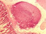
- Lymphatic nodules are present in the mucosa.
Histology
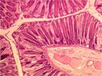
- The mucosa of the large intestine is smooth; there are no villi or microvilli.
- Mucosal glands are much longer and straighter.
- The number of goblet cells in the mucosa is increased compared to the small intestine.
- Mucus is very important for lubrication of the ingesta as it passes through the intestine, particularly as more water is absorbed from the lumen making it drier.
- There are numerous scattered lymph nodules.
- The number of lymph nodules increases compared to the small intestine.
- The submucosa is much reduced in thickness.
- Taenia may be present.
- These are concentrations of the longitudinal muscle layer into long bands.
- When the taenia contract, they cause shortening of the large intestine, which produces saccualtions, or haustra.
- Haustra help to mix the content and to slow the transit time
- Many glands are present in the mucosa and skin of the anal region.
Species Differences
Carnivore
- Digestion is mainly completed by the time chyme enters the large intestine.
- The dog and cat posses two anal sacs.
Ruminant
- Digestion is mainly completed by the time chyme enters the large intestine.
Horse
- Most digestion occurs in the large intestine.
- Taenia present.
Pig
- Taenia present

