Difference between revisions of "Total protein"
Charliecoles (talk | contribs) |
Charliecoles (talk | contribs) |
||
| Line 100: | Line 100: | ||
[[File:NWL 2016 Labfacts Electrophoretogram.png|frameless|330x330px]] | [[File:NWL 2016 Labfacts Electrophoretogram.png|frameless|330x330px]] | ||
| − | |||
| − | |||
| − | |||
| − | |||
| − | |||
| − | |||
| − | |||
| − | |||
| + | [[File:NWL_2016_Labfacts_Electrophoretogram_2.png|alt=|frameless|362x362px]] | ||
{| | {| | ||
Revision as of 10:27, 17 March 2022
Total protein in normal reptiles generally varies between 30 and 80g/l.Hypoproteinaemia is often associated with malnutrition. Other causes include malabsorption, maldigestion (e.g. intestinal parasitism), protein losing enteropathies, severe blood loss and chronic hepatic or renal disease.
Total Protein, Albumin and Globulins
Serum proteins vary widely in their size, structure and function. Abnormal levels of proteins are termed dysproteinaemias. Total protein and albumin concentrations are determined and the globulin concentration arrived at by subtraction. Total protein levels are affected by physiological as well as pathological factors. Total protein levels are low in neonates rising to adult levels by 6 months to 1 year of age. Serum total protein levels are approximately 5% less than those of plasma due to the loss of fibrinogen in the clotting process.
Causes of Hyperproteinaemia in Small Animals
Hyperalbuminaemia
- Dehydration
Hyperglobulinaemia
- Inflammation
- Immune-mediated disease
- Neoplasia
Causes of hypoproteinaemia in Small Animals
Hypoalbuminaemia
- Hepatic insufficiency
- Protein-losing enteropathy
- Protein-losing nephropathy
- Haemorrhage
- Protein malnutrition/malabsorption/maldigestion
- Exudation (body cavity, skin)
- Compensatory for a hyperglobulinaemia
Hypoglobulinaemia
- Protein-losing enteropathy
- Haemorrhage
- Neonates
- Congenital immunodeficiency
Complementary tests in Small Animals
Serum protein electrophoresis, urine protein electrophoresis, radial immunodiffusion for canine IgG, IgA and IgM (suspected immunodeficiency and classification of myelomas).
Causes of hyperproteinaemia in Equine
Hyperalbuminaemia
- Dehydration
Hyperglobulinaemia
- Inflammation
- Immune response to infection
- Neoplasia for example lymphoma (rare)
- Cyathostomiasis, large strongylosis, mixed helminthiasis
Causes of hyproteinaemia in Equine
Hypoalbuminaemia
- Intestinal lymphoma
- Cyathostomiasis, large strongylosis, mixed helminthiasis
- Protein-losing enteropathy
- Advanced hepatic insufficiency – usually fibrosis (Ragwort poisoning)
- Idiopathic granulomatous enteritis
- Salmonellosis
- Clostridiosis
- Protein-losing nephropathy
- Compensatory for a hyperglobulinaemia
- NSAIDS
- Glomerulonephritis/pyelonephritis
Less common causes of hypoabuminaemia
- Starvation
- Chronic hepatitis
- Hepatic neoplasia
- Amyloidosis
- Chronic eosinophilic enteritis (rare)
Hypoglobulinaemia
- Inadequate transfer of colostrum (neonates)
- Severe combined immunodeficiency disease in Arabian foals
Complementary tests in Equine
Protein electrophoresis.
Protein Electrophoresis
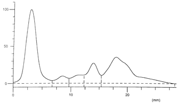
Protein electrophoresis may be indicated when globulins are elevated or there are changes in the albumin to globulin ratio. This is a technique by which the serum proteins are separated into four fractions: albumin, α globulins, β globulins and γ globulins. Canine, feline and equine alpha and beta globulins are further subdivided into α1, α2, β1 and β2 fractions.
α-globulin. Predominantly synthesised in the liver, (α1-fetoprotein synthesised by foetal live cells). α1-globulin fraction includes high density lipoproteins and acute phase proteins, which are inflammatory markers (α1-antitrypsin, α1-acid glycoprotein). The α2-globulin fraction includes very low density lipoproteins, low density lipoproteins (on cellulose acetate) and acute phase proteins (α2-macroglobulin, ceruloplasmin, haptoglobulin).
β-globulin. In most domestic animals except ruminants these can be divided into β1 and β2 -globulins. These include important acute phase proteins such as complement (C3, C4), protein C (a natural anticoagulant in plasma), C-reactive protein, ferritin and amyloid A. The β1-globulin fraction includes some of the low density lipoproteins. Fibrinogen, another acute phase protein slightly trails the β2-globulins. The immunoglobulins IgM and IgA extend from the β2 to the γ region. In response to the antigenic stimulus of some infectious agents, or in plasma cell malignancies, immunoglobulins can be recognised in the β2 zone as well as the γ regions.
γ -globulin. In most animals these are seen as two fractions, γ1 and γ2. IgA, IgM and IgE are mainly found in the γ1 region and IgG mainly in the γ2 region.
Small animals. The details shown in the table below provide information for the interpretation of small animal protein electrophoresis. There is currently a great deal of interest in quantitative measurement of individual acute phase proteins, such as serum amyloid A, C-reactive protein and α-1-acid glycoprotein to assist in the diagnosis of non-specific acute inflammatory disease and conditions such as FIP.
Electrophoretogram
| Index | Band | Rel. Area | Conc. (g/l) |
|---|---|---|---|
| 1 | Albumin | 43.84% | 35.07 |
| 2 | Alpha 1 | 4.12% | 3.30 |
| 3 | Alpha 2 | 5.18% | 4.15 |
| 4 | Beta | 11.28% | 9.02 |
| 5 | Gamma | 35.58% | 28.46 |
| Total | 80.00 |
Interpretation of dysproteinaemias based on the A:G ratio and the protein electrophoresis profile (Kaneko, Jiro J et al. 2008; Parry, BW (ed.) 2008).
| A:G Ratio | Abnormalities | Causes |
|---|---|---|
| Normal (normal electrophoresis trace) | Hyperproteinaemia | Dehydration |
| Hypoproteinaemia | Overhydration Acute blood loss
External plasma loss: extravasation from burns, abrasions, exudative lesions, exudative dermatopathies, external parasites, gastrointestinal disease, diarrhoea. Internal plasma loss: gastrointestinal disease, internal parasites | |
| Decreased | Hypoalbuminaemia | Selective loss of albumin: glomerulonephropathies, nephrosis, nephrotic syndrome, gastrointestinal disease, internal parasites.
Decreased synthesis of albumin: chronic liver disease, malnutrition, chronic inflammatory disease |
| Increased α1-globulins | Acute inflammatory disease: α1-antitrypsin, α1-acid glycoprotein (FIP, FIV) | |
| Increased α2–globulins | Acute inflammatory disease: α2-macroglobulin, ceruloplasmin, haptoglobin
Severe active hepatitis: α2-macroglobulin Acute nephritis: α2-macroglobulin Nephrotic syndrome: α2 -macroglobulin, α2 -lipoprotein (VLDL) | |
| Increased β-globulin | Acute hepatitis: transferrin, hemopexin
Nephrotic syndrome: β2-lipoprotein (LDL), transferrin Suppurative dermatopathies: IgM, C3 | |
| β-γ bridging | Chronic active hepatitis: IgA, IgM | |
| Increased γ-globulin (broad increases)
- polyclonal gammopathies (broad increases): IgG, IgM, IgA |
Chronic inflammatory disease, infectious disease, collagen disease
Chronic hepatitis Hepatic abscess Suppurative disease: feline infectious dermatitis, suppurative dermatitis, tuberculosis Immune-mediated disease: immune-mediated haemolytic anaemia and thrombocytopaenia, Aleutian disease of mink, equine infectious anaemia, systemic lupus erythematosus, autoimmune polyarthritis, autoimmune glomerulonephritis, autoimmune dermatitis, allergies Lymphoma | |
| Increased γ-globulin (sharp increases)
- monoclonal gammopathies (sharp increases): IgG, IgM, IgA |
Lymphoma
Plasma cell dyscrasias: multiple myeloma, Aleutian disease of mink Macroglobulinaemia Benign, nonpathologic | |
| Increased | Increased albumin | Dehydration |
| Decreased globulins | Precolostral neonate
Combined immunodeficiency of Arabian foals Aglobulinaemia |
Equine. Much of the information in the previous table also applies to horses but the α1-globulin fraction has no known significance. Common causes of elevations in the other fractions are:
α2-globulin. High levels are associated with tissue damage such as infection and abscess formation. In the latter the γ-globulins may also be elevated.
β1-globulin. High levels have been associated with intestinal larval pathology. However this is not a consistent finding in all cases and can be normal even when larvae are present.
β2-globulin. High levels can be due to intestinal larval pathology as well as some cases of hepatopathy. In the latter changes would be expected to occur in serum liver enzymes. Monoclonal peaks can occur in horses with lymphoma or plasma cell myeloma.
γ -globulin. Because this includes IgG, rises are seen with antibody responses to infection. Monoclonal peaks can occur in horses with lymphoma or plasma cell myeloma.
Test Codes - Please visit www.nwlabs.co.uk or see our current price list for more information
References
Total Protein, Albumin and Globulins References: NationWide Laboratories

