Difference between revisions of "Fasciola hepatica"
(table) |
|||
| Line 38: | Line 38: | ||
| '''F. Hepatica''' | | '''F. Hepatica''' | ||
|} | |} | ||
| − | |||
| − | |||
| − | |||
| − | |||
| − | |||
| − | |||
| − | |||
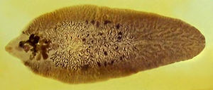
[[Image:Fasciola hepatica.jpg|300px|thumb|right|'''Fasciola hepatica (Copyright Adam Cuerden, Wikimedia Commons) ''']] | [[Image:Fasciola hepatica.jpg|300px|thumb|right|'''Fasciola hepatica (Copyright Adam Cuerden, Wikimedia Commons) ''']] | ||
| Line 53: | Line 46: | ||
===Acute Fascioliasis=== | ===Acute Fascioliasis=== | ||
| − | The acute disease is a less common type of Fasciola hepatica, and generally occurs 2-6 weeks after large ingestion of metacercariae. The young liver flukes migrate through the liver parenchyma causing severe haemorrhaging, due to the damage to | + | The acute disease is a less common type of Fasciola hepatica, and generally occurs 2-6 weeks after large ingestion of metacercariae. The young liver flukes migrate through the liver parenchyma causing severe haemorrhaging, due to the damage to the [[Liver - Anatomy & Physiology|liver]] vasculature. |
| − | + | ||
| − | This occurs in late autumn and winter, mainly between the months of August to October. | + | This occurs in late autumn and winter, mainly between the months of August to October. Outbreaks of acute fascioliasis usually present as sudden deaths. On examination infected animals are weak, with pale mucous membranes. They may also have enlarged livers, and the liver surface may be cover with a fibrinous peritonitis, particularly evident on the ventral lobe. Tracts become filled with blood and degenerate hepatocytes later infiltrated with [[Eosinophils|eosinophils]], [[Lymphocytes|lymphocytes]] and replaced by fibrosis. |
| − | |||
| − | |||
| − | |||
| − | |||
| − | |||
| − | |||
| − | |||
| − | |||
| − | |||
[[Image:Fasciola hepatica - life cycle.jpg|300px|thumb|right|'''Fasciola hepatica life cycle(Copyright Centres for Disease Control and Prevention) ''']] | [[Image:Fasciola hepatica - life cycle.jpg|300px|thumb|right|'''Fasciola hepatica life cycle(Copyright Centres for Disease Control and Prevention) ''']] | ||
| − | + | ||
| + | ===Subactute Fascioliasis=== | ||
| + | This is caused by ingestion of metacercariae over a longer period of time. Some may have migrated to the bile ducts, causing [[cholangitis]] | ||
| + | |||
| + | ===Chronic Fascioliasis=== | ||
| + | |||
======Gross====== | ======Gross====== | ||
*[[Liver - Anatomy & Physiology|liver]] is reduced in size, unevenly | *[[Liver - Anatomy & Physiology|liver]] is reduced in size, unevenly | ||
Revision as of 10:04, 6 July 2010
| Also known as: | Liver Fluke |
Fasciola Hepatica is an hepatic parasite found in mainly in ruminants, namely cows, sheep and goats, but also known to affect horses and pigs. It is commonly found within the UK, with its prevalence ever increasing. It is responsible for a 10-15% production loss in each infected animal, as it affects meat, milk and wool production, so is of huge economic consequence.
Fasiola Hepatica has a definitive ruminant mammalian host and an intermediate molluscian host. The full life cycle is illustrated in the image aside.
| Kingdom | Animalia |
| Phylum | Platyhelminthes |
| Class | Trematoda |
| Subclass | Digenea |
| Order | Echinostomida |
| Family | Fasciolidae |
| Genus | Fasciola |
| Species | F. Hepatica |
Pathogenesis
The severity of the infection is mainly dependent on the number of metacercariae ingested. The Pathogenesis is often described as two-fold. The first stage occuring when the parasite migrates through the liver parenchyma, causing liver damage and haemorrhage. The second phase occurs when the parasite is in the bile ducts, and damage is a result of the haematophagic activity of the adult flukes.
Acute Fascioliasis
The acute disease is a less common type of Fasciola hepatica, and generally occurs 2-6 weeks after large ingestion of metacercariae. The young liver flukes migrate through the liver parenchyma causing severe haemorrhaging, due to the damage to the liver vasculature.
This occurs in late autumn and winter, mainly between the months of August to October. Outbreaks of acute fascioliasis usually present as sudden deaths. On examination infected animals are weak, with pale mucous membranes. They may also have enlarged livers, and the liver surface may be cover with a fibrinous peritonitis, particularly evident on the ventral lobe. Tracts become filled with blood and degenerate hepatocytes later infiltrated with eosinophils, lymphocytes and replaced by fibrosis.
Subactute Fascioliasis
This is caused by ingestion of metacercariae over a longer period of time. Some may have migrated to the bile ducts, causing cholangitis
Chronic Fascioliasis
Gross
- liver is reduced in size, unevenly
- left lobe is most severely affected with atrophy of the extremities
- hypertrophy may occur in some cases
- dorsal lobe
- this changes size and distorts shape of liver
- the surface will be uneven with areas of fibrous tissue replacing the cells damaged by the migrating flukes
- bile ducts
- prominent thick protruding white bile ducts on the visceral surface spreading from the hilus to the left lobe
- the bile ducts are dilated, black, and calcified on cut surface
- numerous adult flukes can be expressed from the bile ducts
- chronic cholangitis
- 'pipe stem' appearance in cattle because bile ducts are very much thickened and often calcified
- bile
- dark brown, thick, and gritty in consistency
NB: the fibrosis which occurs in the chronic stage is realted only partly to the healing of the migratory tracts and the rest may be related to the development of immunity and rechallenge
Microscopically
- reactive hyperplasia of the bile ducts
- substantial inflammatory cell infiltrate and peripheral fibrosis
- calcification of the chronically damaged tissue
