Difference between revisions of "Black Leg"
| Line 1: | Line 1: | ||
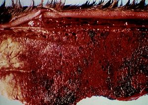
| − | [[Image:Black leg myositis.jpg|right|thumb| | + | [[Image:Black leg myositis.jpg|right|thumb|300px|<small><center>Blackleg myositis (Image sourced from Bristol Biomed Image Archive with permission)</center></small>]] |
{| cellpadding="10" cellspacing="0" border="1" | {| cellpadding="10" cellspacing="0" border="1" | ||
Revision as of 21:36, 18 July 2010
| Also known as: | Blackquarter
|
Description
A bacterial disease affecting cattle and sheep caused by Clostridium chauvoei. Latent spores of this organism are deposited in the muscle and liver of ruminants via the circulation resulting in oedematous and crepitant swelling of the muscles.
Signalment
In cattle it is typically beef breeds who are affected particularly animals in good health and good body condition. More frequently occurs in cattle between 6-24 months old but can affect animals of any age. In some animals lesions occur following muscle trauma, which is thought to activate latent spores in the muscle. In sheep, cases typically occur following some form of injury such as shearing cuts, docking or castration.
Diagnosis
Diagnosis is made on clinical signs and muscle biopsy. Affected muscle is black, dry and infiltrated with small bubbles. The lesions can be present in any muscle. Often in sheep, lesions are deep and quite small. Suspected cases can be confirmed using demonstration of C. chauvoei in affected muscle using the fluorescent antibody test.
History and Clinical Signs
Spores gain entry to GI tract -> blood -> muscle -> lie latent. Under right conditions (usually anaerobic following injury) they germinate and bacilli grow. Toxin damages capillaries -> serosanguinous exudate. Muscle necrosis due to gas producing bacteria -> emphysaema and crepitus The bacteria can cause rapid toxaemia resulting in death, however if clinical signs do occur these can include toxaemia, pyrexia, depression, pulmonary oedema, circulatory collapse lameness and swollen hot muscles later becoming cool as necrosis occurs.
- Pathogenesis:
- Spores gain entry to GI tract -> blood -> muscle -> lie latent. Under right conditions (usually anaerobic following injury) they germinate and bacilli grow. Toxin damages capillaries -> serosanguinous exudate. Muscle necrosis due to gas producing bacteria -> emphysaema and crepitus
- Grossly:
- Early stages
- At muscle periphery
- Dark red
- Distended by serous or serosanguinous exudate
- Wet cut surface
- Old stages
- Centre of lesion is full of gas bubbles, porous, dry, reddish black
- Rancid odour
- Early stages
- Histologically:
- Early stages
- Separation of myofibres by exudate
- Coagulative necrosis
- No nuclei
- Old stage
- Fragmented muscle fibres separated by gas bubbles
- Gram positive bacilli may be found in clumps
- Early stages
