Difference between revisions of "Gingiva"
| Line 8: | Line 8: | ||
===Labiogingival groove=== | ===Labiogingival groove=== | ||
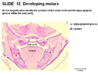
| − | [[Image:Labiogingival Groove Histology.jpg|thumb|right| | + | [[Image:Labiogingival Groove Histology.jpg|thumb|right|200px|Labiogingival Groove Histology - Copyright RVC 2008]] |
The '''labiogingival groove''' is the junction between the '''labial border''' and '''gingival line''' on the distal/medial surface of the incisor teeth. | The '''labiogingival groove''' is the junction between the '''labial border''' and '''gingival line''' on the distal/medial surface of the incisor teeth. | ||
Revision as of 11:24, 9 September 2010
Introduction
Gingiva wrap around the neck of each tooth forming the gums. The gums are useful clinically in assessing health status of an animal.
Structure and Function of the Gingiva
Gingiva is mucosal tissue over alveolar bone. It has a stratified squamous epithelium, with some keratinisation. It resists friction of food during mastication by being tightly bound to the underlying bone. It recedes with age, exposing the cervical part of the tooth. It is usually salmon pink in healthy animals. A colour change indicates pathology.
Labiogingival groove
The labiogingival groove is the junction between the labial border and gingival line on the distal/medial surface of the incisor teeth.
Vasculature and Innervation of the Gingiva
The gingiva is supplied by the superior and inferior alveolar arteries.
There are blood vessels in the dental pulp cavity and a single branch in each major elevation of the crown.
Innervation is from the trigeminal nerve (CN V).
Species Differences
Canine
Some breeds of dog have dark gums, e.g. chow chow.
