Difference between revisions of "Small Animal Dermatology Q&A 11"
Ggaitskell (talk | contribs) |
Ggaitskell (talk | contribs) |
||
| Line 1: | Line 1: | ||
{{Template:Manson Moriello}} | {{Template:Manson Moriello}} | ||
| − | [[Image:Small Animal Dermatology Q&A | + | [[Image:Small Animal Dermatology Q&A 11a.jpg|centre|500px]]<br> |
<br /> | <br /> | ||
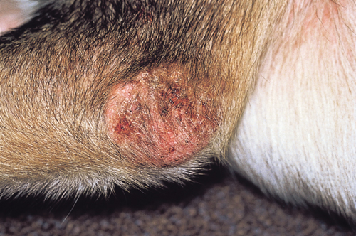
| − | ''' | + | '''An intensely pruritic dog was presented for examination. The dog’s pruritus developed acutely approximately 3 weeks ago. The dog had no history of skin disease prior to this episode. The dog was normal on physical examination except for being intensely pruritic and having ‘scaly’ elbows. The organism shown was found on a skin scraping from the elbow of the patient.''' |
<br /> | <br /> | ||
<FlashCard questions="3"> | <FlashCard questions="3"> | ||
| − | |q1=What is the | + | |q1=What is the organism? |
|a1= | |a1= | ||
| − | + | ''Sarcoptes scabiei'' mite. | |
| − | |||
| − | |||
|l1= | |l1= | ||
| − | |q2=What are | + | |q2=What clinical signs are associated with this parasite infestation? |
|a2= | |a2= | ||
| − | The | + | This is a highly contagious mite that causes intense pruritus. The history of an acute onset of intense pruritus is common. The mites burrow in thinly haired areas, and intense ventral pruritus may be the first clinical sign noted. In many cases, there is a history of exposure to affected dogs or high-risk exposure situations (e.g. stray dogs, boarding kennel, visit to a grooming facility, visit to a park). In many patients, lesions may be absent but thinly haired areas, the ventrum, elbows, and ear margins, are often good sites to find mites. Both deep and superficial skin scrapings should be done to increase the chances of finding the mites. |
| − | |||
| − | |||
| − | |||
| − | |||
| − | |||
| − | |||
| − | |||
| − | |||
| − | |||
|l2= | |l2= | ||
| − | |q3=What are the | + | |q3=What new diagnostic test is available, and what are the limitations of the test? |
|a3= | |a3= | ||
| − | + | Recently, an in vitro serum antibody test was marketed in Sweden for the diagnosis of this parasite. The test is reported to have a sensitivity of 83% and a specificity of 92% (Curtis, 2001). | |
| − | |||
| − | |||
|l3= | |l3= | ||
</FlashCard> | </FlashCard> | ||
Revision as of 10:15, 6 June 2011
| This question was provided by Manson Publishing as part of the OVAL Project. See more small animal dermatological questions |
An intensely pruritic dog was presented for examination. The dog’s pruritus developed acutely approximately 3 weeks ago. The dog had no history of skin disease prior to this episode. The dog was normal on physical examination except for being intensely pruritic and having ‘scaly’ elbows. The organism shown was found on a skin scraping from the elbow of the patient.
| Question | Answer | Article | |
| What is the organism? | Sarcoptes scabiei mite. |
[[|Link to Article]] | |
| What clinical signs are associated with this parasite infestation? | This is a highly contagious mite that causes intense pruritus. The history of an acute onset of intense pruritus is common. The mites burrow in thinly haired areas, and intense ventral pruritus may be the first clinical sign noted. In many cases, there is a history of exposure to affected dogs or high-risk exposure situations (e.g. stray dogs, boarding kennel, visit to a grooming facility, visit to a park). In many patients, lesions may be absent but thinly haired areas, the ventrum, elbows, and ear margins, are often good sites to find mites. Both deep and superficial skin scrapings should be done to increase the chances of finding the mites. |
[[|Link to Article]] | |
| What new diagnostic test is available, and what are the limitations of the test? | Recently, an in vitro serum antibody test was marketed in Sweden for the diagnosis of this parasite. The test is reported to have a sensitivity of 83% and a specificity of 92% (Curtis, 2001). |
[[|Link to Article]] | |
