Difference between revisions of "Avian Medicine Q&A 17"
(Created page with "<br style="clear:both;" /> {| align="left" width="100%" style="background-color:#04B4AE" |- | align="center" | 90px|Mansonlogo | align="left" | This ques...") |
|||
| Line 3: | Line 3: | ||
|- | |- | ||
| align="center" | [[File:Manson_logo.gif|90px|Mansonlogo]] | | align="center" | [[File:Manson_logo.gif|90px|Mansonlogo]] | ||
| − | | align="left" | This question was provided by [[:Category:Manson|Manson Publishing]] as part of the [[OVAL Project]]. See more [[Category: Avian Medicine Q&A|Avian Medicine questions]] | + | | align="left" | This question was provided by [[:Category:Manson|Manson Publishing]] as part of the [[OVAL Project]]. See more [[:Category: Avian Medicine Q&A|Avian Medicine questions]] |
|} | |} | ||
<br><br><br> | <br><br><br> | ||
Revision as of 21:33, 2 August 2011
| This question was provided by Manson Publishing as part of the OVAL Project. See more Avian Medicine questions |
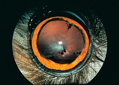
The condition in the right eye of this long-eared owl(Asio otus) is illustrated using mydriasis induced by air sac perfusion anaesthesia (APA).
| Question | Answer | Article | |
| What is the condition and aetiology responsible for the ocular discharge? | This is post-traumatic subluxation of the lens with rupture of the lens capsule and zonula fibres in the ventronasal lens periphery with consecutive coloboma of the lens and iridodonesis.
There is traumatic nuclear cataract and false cataract with multiple black pigment spots on the anterior lens capsule with slight dyscoria of the iris. There is a dim reddish fundus reflex due to the absence of a tapetum lucidum and the poorly pigmented fundus in nocturnal bird species. |
[[|Link to Article]] | |
| Which additional ophthalmological diagnostic procedures would you perform? | The slight dyscoria of the iris as well as the black spots on the anterior lens capsule are residues of the posterior surface of the iris due to partial dissolution of the posterior synechia.
Measure the intraocular pressure immediately – physiological value 10.2 ± 2.1 mmHg – preferably using an electronic tonometer. This may initially be slightly decreased due to uveitis but may be increased by up to 40 mmHg, indicative of glaucoma. Since this is a condition with a traumatic aetiology, ophthalmoscopy of the contralateral eye – which usually appears outwardly healthy – is indicated for prompt recognition of intravitreal haemorrhage. |
[[|Link to Article]] | |
| What is the treatment? | Rupture of the lens capsule leads to lens protein leakage into the anterior chamber; upon contact with the anterior uvea this is recognized as foreign material, resulting in uveitis.
Cataract resection – preferably by phacoemulsification – is obligatory to prevent complete inflammatory destruction of the eye. |
[[|Link to Article]] | |
