Difference between revisions of "Immunodeficiencies - WikiBlood"
m (Text replace - 'Neutrophils - WikiBlood' to 'Neutrophils') |
m (Text replace - 'Lymphocytes - WikiBlood' to 'Lymphocytes') |
||
| Line 22: | Line 22: | ||
*Primary immunodeficiencies may affect either the [[Innate Immune System - WikiBlood|innate immune system]] or the [[Adaptive Immune System - WikiBlood|adaptive immune system]] | *Primary immunodeficiencies may affect either the [[Innate Immune System - WikiBlood|innate immune system]] or the [[Adaptive Immune System - WikiBlood|adaptive immune system]] | ||
*They are categorised by either the type or the developmental stage of the cells involved | *They are categorised by either the type or the developmental stage of the cells involved | ||
| − | *Lymphoid cell disorders affect [[Lymphocytes | + | *Lymphoid cell disorders affect [[Lymphocytes#T cells|T cells]] or [[Lymphocytes#B cells|B cells]] (or both) |
*Myeloid cell disorders affect phagocytic function | *Myeloid cell disorders affect phagocytic function | ||
*The severity of the immunodeficiency depends on at which stage in development the problem occurs | *The severity of the immunodeficiency depends on at which stage in development the problem occurs | ||
**E.g. Defects early on in development will affect the entire immune system | **E.g. Defects early on in development will affect the entire immune system | ||
| − | *[[Lymphocytes | + | *[[Lymphocytes#T cells|T cell]] deficiencies can affect both the cell-mediated and humoral response as [[Lymphocytes#T cells|T cells]] play a central role in the immune system |
===Deficiencies of Innate Immunity=== | ===Deficiencies of Innate Immunity=== | ||
| Line 60: | Line 60: | ||
*Occurs in 2-3% of Arabian foals | *Occurs in 2-3% of Arabian foals | ||
*Defect in DNA-dependent protein kinase gene | *Defect in DNA-dependent protein kinase gene | ||
| − | **Gene codes for a DNA repair enzyme involved in V(D)J recombination for antigen receptors of [[Lymphocytes | + | **Gene codes for a DNA repair enzyme involved in V(D)J recombination for antigen receptors of [[Lymphocytes|lymphocytes]] (e.g. Ig and TCR) |
| − | *No functional [[Lymphocytes | + | *No functional [[Lymphocytes#B cells|B cells]] or [[Lymphocytes#T cells|T cells]] |
*Foals develop infections (usually around 8 weeks of age as maternal [[Immunoglobulins - WikiBlood|antibody]] in [[Materno-fetal immunity - WikiBlood#Passive transfer via colostrum|colostrum]] wanes around this time) | *Foals develop infections (usually around 8 weeks of age as maternal [[Immunoglobulins - WikiBlood|antibody]] in [[Materno-fetal immunity - WikiBlood#Passive transfer via colostrum|colostrum]] wanes around this time) | ||
*Foals usually die from bronchopneumonia | *Foals usually die from bronchopneumonia | ||
| Line 68: | Line 68: | ||
*Affects Basset Hounds and Corgis | *Affects Basset Hounds and Corgis | ||
*X-linked recessive defect in the gene coding for the IL-2 receptor | *X-linked recessive defect in the gene coding for the IL-2 receptor | ||
| − | **IL-2 receptor is a receptor for the cytokine IL-2 which causes [[Lymphocytes | + | **IL-2 receptor is a receptor for the cytokine IL-2 which causes [[Lymphocytes#T cells|T cells]] to proliferate |
*Causes lymphoid hypoplasia, stunted growth and increases the animal's susceptibility to infection | *Causes lymphoid hypoplasia, stunted growth and increases the animal's susceptibility to infection | ||
*Animal usually dies from pneumonia or sepsis as the level of maternal [[Immunoglobulins - WikiBlood|antibody]] decreases | *Animal usually dies from pneumonia or sepsis as the level of maternal [[Immunoglobulins - WikiBlood|antibody]] decreases | ||
| Line 89: | Line 89: | ||
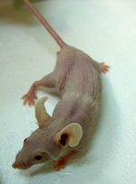
[[Image:Nude Mouse.jpg|right|thumb|150px|Nude Mouse - Copyright Michigan State University- Carcinogenesis Laboratory]] | [[Image:Nude Mouse.jpg|right|thumb|150px|Nude Mouse - Copyright Michigan State University- Carcinogenesis Laboratory]] | ||
*Severe Combined Immune Deficiency(SCID) | *Severe Combined Immune Deficiency(SCID) | ||
| − | **No functional [[Lymphocytes | + | **No functional [[Lymphocytes#B cells|B cells]] or [[Lymphocytes#T cells|T cells]] |
*Athymic nude mice (no [[Thymus - Anatomy & Physiology|thymus]]) | *Athymic nude mice (no [[Thymus - Anatomy & Physiology|thymus]]) | ||
| − | **No functional [[Lymphocytes | + | **No functional [[Lymphocytes#T cells|T cells]] |
**Cell-mediated immunodeficiency | **Cell-mediated immunodeficiency | ||
| Line 110: | Line 110: | ||
[[Image:Kinetics of FeLV 2.jpg|thumb|right|150px|Kinetics of FeLV - Copyright Dr Brian Catchpole BVetMed PhD MRCVS]] | [[Image:Kinetics of FeLV 2.jpg|thumb|right|150px|Kinetics of FeLV - Copyright Dr Brian Catchpole BVetMed PhD MRCVS]] | ||
*Oncogenic retrovirus | *Oncogenic retrovirus | ||
| − | *Causes neoplasia (lymphoma), myelosuppression (anaemia) and immunosuppression (of [[Lymphocytes | + | *Causes neoplasia (lymphoma), myelosuppression (anaemia) and immunosuppression (of [[Lymphocytes#T cells|T cells]]) |
*2 strains: | *2 strains: | ||
**FeLV-A | **FeLV-A | ||
| Line 143: | Line 143: | ||
*Subtypes A, B and D | *Subtypes A, B and D | ||
*Causes increased susceptibility to infections and neoplasia | *Causes increased susceptibility to infections and neoplasia | ||
| − | *Specifically destroys [[Lymphocytes | + | *Specifically destroys [[Lymphocytes#Helper CD4+|CD4+ T cells]] |
*Virus is present in saliva, blood and other bodily fluids | *Virus is present in saliva, blood and other bodily fluids | ||
*Feral and outdoor cats (mostly tom cats) are most at risk | *Feral and outdoor cats (mostly tom cats) are most at risk | ||
Revision as of 12:37, 12 June 2010
| This article has been peer reviewed but is awaiting expert review. If you would like to help with this, please see more information about expert reviewing. |
|
|
Introduction
Like any system in the body the immune system can go wrong. Autoimmunity is when the immune system begins to attack itself. Immunodeficiency is when the immune system fails to protect itself from disease.
If the immunodeficient defect is present at birth and is therefore a result of a genetic or developmental abnormality, it is called a primary immunodeficiency.
Secondary immunodeficiency, sometimes called acquired immunodeficiency, is the loss of immune function during life, caused by exposure to harmful agents.
Immunodeficiencies can be treated by the replacement of the defective or missing protein, cells or gene. However, in veterinary medicine, vaccinations and drugs are the most common treatments for immunodeficiency.
Primary Immunodeficiency
- Primary immunodeficiencies may affect either the innate immune system or the adaptive immune system
- They are categorised by either the type or the developmental stage of the cells involved
- Lymphoid cell disorders affect T cells or B cells (or both)
- Myeloid cell disorders affect phagocytic function
- The severity of the immunodeficiency depends on at which stage in development the problem occurs
- E.g. Defects early on in development will affect the entire immune system
- T cell deficiencies can affect both the cell-mediated and humoral response as T cells play a central role in the immune system
Deficiencies of Innate Immunity
Canine Cyclic Haematopoiesis
- Also called Grey Collie Syndrome
- Autosomal recessive
- Insertion mutation in AP3B1 gene
- Diluted grey coat colour, stunted growth, poor wound healing
- Neutropenia every 2 weeks which lasts 3-4 days due to cyclic production of cells from bone marrow
- Animals are prone to recurrent infections, mainly from the respiratory and gastrointestinal tract
- E.g. pyrexia, diarrhoea, gingivitis and arthritis
- Puppies can be distinguished from other litter mates by the diluted grey colouring
- Affected puppies show symptoms such as fever, joint pain and eye, skin and respiratory infections from 8 weeks of age
- Affected animals rarely live beyond 2-3 years with most puppies dying within a few weeks of birth
Canine Leukocyte Adhesion Deficiency (CLAD)
- Occurs in Irish Setters
- Missence mutation of -Cys-36-Ser- in CD18 molecule
- CD18 is required for neutrophil migration and phagocytosis
- Recurrent bacterial infection
- Neutrophilia (neutrophils remain in the blood and are unable to fight infection in the tissue)
Bovine Leukocyte Adhesion Deficiency (BLAD)
- Occurs in Holstein cattle
- Missence mutation of -Asp-128-Gly in CD18 molecule
- Recurrent infection, e.g. pneumonia
Deficiencies of Adaptive Immunity
Equine Severe Combined Immune Deficiency (Equine SCID)
- Autosomal recessive
- Occurs in 2-3% of Arabian foals
- Defect in DNA-dependent protein kinase gene
- Gene codes for a DNA repair enzyme involved in V(D)J recombination for antigen receptors of lymphocytes (e.g. Ig and TCR)
- No functional B cells or T cells
- Foals develop infections (usually around 8 weeks of age as maternal antibody in colostrum wanes around this time)
- Foals usually die from bronchopneumonia
Canine X-Linked Severe Combined Immune Deficiency (Canine SCID)
- Affects Basset Hounds and Corgis
- X-linked recessive defect in the gene coding for the IL-2 receptor
- IL-2 receptor is a receptor for the cytokine IL-2 which causes T cells to proliferate
- Causes lymphoid hypoplasia, stunted growth and increases the animal's susceptibility to infection
- Animal usually dies from pneumonia or sepsis as the level of maternal antibody decreases
Selective IgA deficiency of German Shepherd Dogs
- Poorly understood
- Linked to other disease syndromes such as deep pyoderma, inflammatory bowel disease, anal furunculosis and disseminated aspergillosis
- IgA deficiency so more susceptible to mucosal disease
Immunodeficiency of Weimaraners, Irish Wolfhounds and Miniature Dachshunds
- Unknown aetiology
- Inherited defects
- Low levels of circulating IgM and IgG
- Impaired neutrophil function
- Causes recurrent pyrexia and infections
Laboratory Examples of Severe Combined Deficiency
- Knock-out mice
- E.g. Gene coding for CD4, CD8, IL-10 removed
Secondary Immunodeficiency
- There are many causes of secondary immunodeficiency
- Most deficiencies are not genetic
- Most are agent-induced, such as from X-ray radiation and immunosuppressive drugs
Viral Causes
Feline Leukaemia Virus (FeLV)

- Oncogenic retrovirus
- Causes neoplasia (lymphoma), myelosuppression (anaemia) and immunosuppression (of T cells)
- 2 strains:
- FeLV-A
- Natural strain
- FeLV-B
- Formed through FeLV-A recombining with endogenous retroviral sequences in the feline genome
- Increases the risks of lymphoma
- FeLV-C
- Formed from the spontaneous mutation of FeLV-A
- Is more myelosuppressive
- FeLV-A
- Virus replicates in the oropharyngeal lymphoid tissue causing a viraemia (virus circulating in the bloodstream) which then spreads to the systemic lymphoid tissue
- Shed in saliva
- Passed by oronasal route, e.g. mutual grooming
- Kittens between 6 weeks and 6 months are most susceptible
- 60% of cats will become immune to the disease and recover
- Cats that are persistently viraemic will progress to develop FeLV-associated diseases
- Some cats will become viraemic again if treated with corticosteroids or stressed if the infection lies dormant in the bone marrow
- Diagnosis:
- ELISA
- Rapid-Immuno-Migration
- Western Blot
- Virus Isolation
- Immunofluorescence
- PCR
- Treatment:
- Antibiotics for secondary infection
- Anti-retroviral therapy
- For vaccinations see here
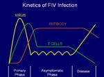
Feline Immunodeficiency Virus (FIV)
- Lentivirus
- Subtypes A, B and D
- Causes increased susceptibility to infections and neoplasia
- Specifically destroys CD4+ T cells
- Virus is present in saliva, blood and other bodily fluids
- Feral and outdoor cats (mostly tom cats) are most at risk
- Virus replicates in lymphoid tissue
- Can remain asymptomatic
- Causes pyrexia and lymphadenopathy
- Transmitted by biting
- Diagnosis:
- ELISA
- Rapid-Immuno-Migration
- Western Blot
- Virus Isolation
- Immunofluorescence
- PCR
- Treatment:
- Antibiotics for secondary infection
- Anti-retroviral therapy
- For vaccinations see here
Bovine Immunodeficiency Virus (BIV)
- Lentivirus (non-oncogenic)
- Causes a persistent viral infection and lymphocytosis
- Immunocompromised cattle may develop secondary infections
- The transmission is not well known, but the following possibilities are being researched:
- Through milk
- Through infected semen (e.g.artificial insemination)
- Placental transfer
- Diagnosis:
- Western Blot
- PCR
Toxic Causes
- Poisons
Iatrogenic Causes
- Drugs
- Corticosteroids
- Cyclosporin
- Cytotoxic cancer therapy
Other Causes
- Malnutrition
- Chronic disease
- Stress
- Senescence
Links
Internal
External
- Grey Collie Syndrome Information on Canine Cyclic Haematopoeisis (Grey Collie Syndrome) including new research into treating the condition and a clinical example
- Nude Mice Information on nude mice and their role in cancer research
Immunodeficiencies Flashcards
References
Books
- Ivan Roitt: Essential Immunology, Ninth edition
- Goldsby, Kindt, & Osbourne KUBY Immunology, Fourth edition
Lecture Notes
- Dr Brian Catchpole BVetMed PhD MRCVS
Websites
- Michelle Tennis & Peggy Melton http://www.bitoheavencollies.com


