Difference between revisions of "Toxoplasmosis - Sheep"
| Line 59: | Line 59: | ||
Apart from ingestion of oocysts in the environment, the only other method of transmission of toxoplasmosis to sheep is vertical spread from mother to foetus during pregnancy. This is because sheep are herbivorous, and do not consume animal tissues containing cysts. The outcome of transplacental infection depends on the stage of pregnancy. Infection in early gestation usually causes foetal death, as the foetal immune system is immature at this stage. In mid-gestation, infection may cause the birth of weak or stillborn lambs, sometimes accompanied by a mummified sibling. Ewes infected in the third trimester normally give birth to infected but clinically normal lambs. | Apart from ingestion of oocysts in the environment, the only other method of transmission of toxoplasmosis to sheep is vertical spread from mother to foetus during pregnancy. This is because sheep are herbivorous, and do not consume animal tissues containing cysts. The outcome of transplacental infection depends on the stage of pregnancy. Infection in early gestation usually causes foetal death, as the foetal immune system is immature at this stage. In mid-gestation, infection may cause the birth of weak or stillborn lambs, sometimes accompanied by a mummified sibling. Ewes infected in the third trimester normally give birth to infected but clinically normal lambs. | ||
| − | |||
| − | |||
| − | |||
| − | |||
| − | |||
| − | |||
| − | |||
| − | |||
| − | |||
| − | |||
| − | |||
| − | |||
| − | |||
| − | |||
| − | |||
| − | |||
| − | |||
| − | |||
| − | |||
| − | |||
| − | |||
| − | |||
| − | |||
| − | |||
| − | |||
| − | |||
| − | |||
| − | |||
| − | |||
| − | |||
| − | |||
| − | |||
| − | |||
| − | |||
| − | |||
| − | |||
| − | |||
| − | |||
| − | |||
| − | |||
| − | |||
| − | |||
| − | |||
| − | |||
| − | |||
| − | |||
| − | |||
| − | |||
| − | |||
| − | |||
| − | |||
| − | |||
| − | |||
| − | |||
| − | |||
| − | |||
| − | |||
| − | |||
| − | |||
| − | |||
| − | |||
| − | |||
| − | |||
| − | |||
| − | |||
| − | |||
| − | |||
| − | |||
| − | |||
| − | |||
| − | |||
| − | |||
| − | |||
| − | |||
| − | |||
| − | |||
| − | |||
| − | |||
| − | |||
| − | |||
| − | |||
| − | |||
| − | |||
| − | |||
| − | |||
| − | |||
| − | |||
| − | |||
| − | |||
| − | |||
| − | |||
| − | |||
| − | |||
| − | |||
| − | |||
| − | |||
| − | |||
| − | |||
| − | |||
==Signalment== | ==Signalment== | ||
Revision as of 15:26, 13 August 2010
| This article is still under construction. |
Description
Toxoplasmosis is the disease caused by Toxoplasma gondii, an intracelluler protozoan parasite. Although the definitive host is the cat, T. gondii can infect all mammals including man and is a significant cause of abortion in sheep and goats. Toxoplasmosis does not seem to cause disease in cattle.
Life Cycle
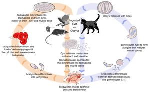
There are three infectious stages of Toxoplasma gondii: 1) sporozoites; 2) actively reproducing tachyzoites; and 3) slowly multiplying bradyzoites. Tachyzoites and bradyzoites are found in tissue cysts, whereas sporozoites are containted within oocysts, which are excreted in the faeces. This means that the protozoa can be transmitted by ingestion of oocyst-contaminated food or water, or by consumption of infected tissue.
In naive cats, Toxoplasma gondii undergoes an enteroepithelial life cycle. Cats ingests intermediate hosts containing tissue cysts, which release bradyzoites in the gastrointestinal tract. The bradyzoites penetrate the small intestinal epithelium and sexual reproductio ensues, eventually resulting the production of oocysts. Oocysts are passed in the cat's faeces and sporulate to become infectious once in the environment. These can then be ingested by other mammals, including sheep.
When sheep ingest oocysts, T.gondii intiates extraintestinal replication. This process is the same for all hosts, and also occurs when carnivores ingest tissue cysts in other animals. Sporozoites (or bradyzoites, if cysts are consumed) are released in the intestine to infect the intestinal epithelium where they replicate. This produces tachyzoites, which reproduce asexually within the infected cell. When the infected cell ruptures, tachyzoites are released and disseminate via blood and lymph to infect other tissues. Tachyzoites then replicate intracellularly again and the process continues until the host becomes immune or dies. If the infected cell does not burst, tachyzoites eventually encyst as bradyzoites and persist for the life of the host. Cyst are most commonly found in the brain or skeletal muscle, and are a source of infection for carnivorous hosts.
Transmission to Sheep
Oocysts in the Environment
Infected cats shed oocysts continuously between days 3 and 14 post-infection. During this time, hundreds of millions of oocysts may be shed. The main sources of feline toxoplasma infection are chronically infected birds and rodents. Rodents are particularly important since they can pass T. gondii infection to their offspring without causing clinical disease. This means that a farm may develop a reservoir of T. gondii tissue cysts with the potential to cause feline infection and massive oocyst excretion. In turn, environments may easily become contaminated with a high oocyst burden when a cat is introduced.
Sheep are often kept in an environment that is significantly contaminated with oocysts, and infection follows ingestion of infected food, primarily contaminated pasture. Fields treated with manure or bedding from buildings to which cats have access result in high levels of ovine toxoplasmosis, and insecure storage of supplementary feeds also poses a risk.
Members of the cat family are the definitive hosts of the parasite and tend to become infected for the first time when they start hunting and eating wild rodents and birds already infected with T. gondii. Following consumption of T. gondii cysts, the parasites excyst in the gut of the cat and invade and infect host cells. Sexual development of the parasite takes place in the gut of the cat resulting in the production of oocysts which are shed in the faeces. Shedding usually occurs around 3–10 days after initial infection and may continue for 2–3 weeks (Dubey and Beattie, 1988). During this period a cat may shed over 100 million oocysts and experimental studies in sheep have shown that a dose of only 200 oocysts may cause abortion in previously naı¨ve pregnant sheep (McColgan, Buxton and Blewett, 1988). The importance of oocysts as a source of infection for sheep, has been supported by studies showing an association with infection and contamination of feed or grazing land with sporulated oocysts (Plant, Richardson and Moyle, 1974; Faull, Clarkson and Winter, 1986) and also work showing an association with cats on farms and prevalence of T. gondii in sheep (Skjerve et al. 1998). Further studies looking at development of specific antibodies in sheep, as an indicator of exposure to T. gondii, have shown that there is an increase in seroprevalence associated with age. This indicates that there is extensive environmental contamination with T. gondii oocysts and that most infections in sheep occur following exposure to the parasite after birth (Waldeland, 1977; Blewett, 1983; Lunden,Nasholm and Uggla, 1994). Recent studies have indicated that there is widespread environmental contamination with T. gondii oocysts (Dabritz et al. 2007).
Congenital Transmission
Apart from ingestion of oocysts in the environment, the only other method of transmission of toxoplasmosis to sheep is vertical spread from mother to foetus during pregnancy. This is because sheep are herbivorous, and do not consume animal tissues containing cysts. The outcome of transplacental infection depends on the stage of pregnancy. Infection in early gestation usually causes foetal death, as the foetal immune system is immature at this stage. In mid-gestation, infection may cause the birth of weak or stillborn lambs, sometimes accompanied by a mummified sibling. Ewes infected in the third trimester normally give birth to infected but clinically normal lambs.
Signalment
Diagnosis
Clinical Signs
- Clinical outbreaks of toxoplasmosis are sporadic
- Immunity is acquired before tupping
- Significant ill-effects are unlikely if immune ewes are infected during pregnancy
- Not shed from sheep to sheep so predicting outbreaks is difficult
Laboratory Tests
Pathology
Aborted ewes show focal necrotic placentitis with white lesions in the cotyledons and foetal tissue
Treatment
- Toxovax vaccine
- Live, avirulent strain of Toxoplasma
- Does not form bradyzoites or tissue cysts
- Killed by host immune system
- Single dose given 6 weeks before tupping
- Protects for 2 years
- Immunity boosted by natural challenge
- Medicated feed can be given daily during the main risk period
- 14 weeks before lambing
- The best method of protection is to prevent cats from contaminating the pasture, lambing sheds and feed stores
The extent of environmental contamination with T. gondii oocysts is thus related to the distribution and behaviour of cats. Measures to reduce environmental contamination by oocysts should be aimed at reducing the number of cats capable of shedding oocysts. This would include attempts to limit their breeding. If male cats are caught, neutered and returned to their colonies the stability ofthe colony is maintained; fertile male cats do not challenge the neutered males12 and breeding is controlled. Thus the maintenance ofa small healthy population of mature cats will reduce oocyst excretion as well as help to control rodents. Sheep feed should be kept covered at all times to prevent its contamination by cat faeces.
Prognosis
Links
References
- Buxton, D (1990) Ovine toxoplasmosis: a review. Journal of the Royal Society of Medicine, 83, 509-511.
- Innes, E A et al (2009) Ovine toxoplasmosis. Parastiology, 136, 1887–1894.
- Buxton, D et all (2007) Toxoplasma gondii and ovine toxoplasmosis: New aspects of an old story. Veterinary Parasitology, 147, 25-28.
- Dubey, J P (2009) Toxoplasmosis in sheep — The last 20 years. Veterinary Parasitology, 163, 1-14.
