Difference between revisions of "Mediastinum - Anatomy & Physiology"
Jump to navigation
Jump to search
| Line 1: | Line 1: | ||
| − | + | ||
| − | |||
| − | |||
| − | |||
| − | |||
| − | |||
| − | |||
| − | |||
| − | |||
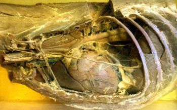
[[Image:Dogthorax1.jpg|right|thumb|350px|''The mediastinum is visible in this dog dissection, left lung removed. ©RVC 2008]] | [[Image:Dogthorax1.jpg|right|thumb|350px|''The mediastinum is visible in this dog dissection, left lung removed. ©RVC 2008]] | ||
| Line 19: | Line 11: | ||
| − | [[Category:Respiratory System]] | + | [[Category:Respiratory System - Anatomy & Physiology]] |
Revision as of 13:03, 10 September 2010
Introduction
The mediastinum divides the thoracic cage into two halves. It extends from the Spine to the Sternum and contains many structures including blood vessels, nerves, oesophagus, trachea and heart.
References
- Sjaastad, O.V., Hove, K. and Sand, O. (2004) Physiology of Domestic Animals. Oslo: Scandinavian Veterinary Press.
