Tooth - Anatomy & Physiology
Anatomy of the Enamel Organ
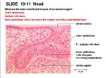
The main components which form the enamel organ are:
- Outer epithelium
- Stellate reticulum- star shaped cells lying between the outer and inner epithelial layers. It has the appearance of connective tissue but is of epithelial derivation.
- Inner epithelium which becomes the enamel secreting ameloblast layer
The enamel organ has many different components. These consist of:
Crown
The crown is covered by enamel. It meets the root at the cemento-enamel junction (CEJ).
The crown of incisors have only one cusp. The crown of molars have up to 4 cusps for the grinding of food.
Root
Teeth may have one or more roots. The furcation angle is the point where roots diverge. The root ends in an apex which is where the nerves, blood vessels and lymphatics travel to the pulp. Hypsodont teeth can have open roots (aradicular) e.g. in rabbits which have continued growth. Hypsodont teeth can have closed roots (radicular) e.g. horse where growth decreases with age. Brachydont teeth have no capacity for growth and so the roots are closed.
Species Differences
The apex has a single foramen in dogs and cats. It remains open in herbivores. In the horse, the apex closes as the animal ages. Brachiocephalic dogs often have fused roots. Equine incisors have fused roots. In the horse's canines, the size of the root is much larger than the crown.
Alveolar Bone
The alveolar processes of the jaw consists of the alveolar bone, trabecular bone and compact bone.
The densest bone called the cribiform plate lines the alveolus. This appears white on radiographs and is referred to as the lamina dura.
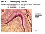
The main cells of the enamel organ are:

