Hoof - Anatomy & Physiology
Introduction
The equine hoof is defined from a physiologic persepctive as the modified skin covering the tip of the digit and all enclosed structures. The hoof provides protection to the distal limb and is formed by keratinisation of the epithelial layer and modification of the underlying dermis. The keratin in the epidermis, when thickened and cornified, is referred to as horn. Horn makes up the outer surface if the hoof and is particularly resistant to mechanical and chemical damage.
Five Segments of the Hoof
Periople Coronary Segment Wall Segment Sole Segment Frog-Bulb Segment
Structure and Function
Equine Hoof
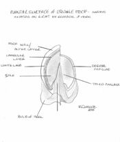
The horny material encasing the distal phalanx is known as the hoof. It is continuous with the skin at the coronary region. The hoof can be divided into three topographical regions; the wall, the frog and the sole. The wall forms the medial, lateral and dorsal aspect of the hoof. It can be further divided into the toe, quarters and heels. At the heel the walls reflect back on themselves at a point called the angles and in doing so forms the bars. The bars fade out before they meet cranially. The frog sits between the bars and has an apex facing dorsally, with 2 crura flanking a central sulcus. Between the crus and bar of each half lies the collateral sulcus. Opposite the apex the frog expands forming the bulbs of the heel. The sole is the area dorsal to the bars and apex of the frog enclosed by the hoof wall. The area where the bars and wall enclose it is known as the angle of the sole.
The dermis of the distal phalanx is arranged in hundreds of leaves or laminae, which have microscopic secondary laminae. The coronary region has a germinative layer associated with papillae that is responsible for producing the horn tubules that make up the hoof wall. This wall glides distally at a rate of 5-6mm a month and by forming epidermal laminae itself it interdigitates with the underlying dermal laminae. Neither of these laminae are pigmented so when the epidermal laminae appear on the solar surface, a non-pigmented region known as the white line appears. The white line is used as important landmark in farriery as structures central to the line will be dermal and so vascular and sensitive. The dermis in the frog is also arranged in papillae and produces incompletely keratinised flexuous horn tubules resulting in a soft, elastic horn. The hypodermis of the region of the frog forms the digital cushion. This lies between the ungual cartilages and is collagenous, elastic tissue infiltrated by adipose tissue. At the bulbs of the heel it is subcutaneous and is soft and loose in texture. The sole area also has papillae that produces superficially flakey horn. The coronary part of the wall is surrounded by a bony prominence called the periople. This soft, lightly coloured area is restricted to this proximal area and is produced by the germative layer covering the papillae. The rest of the hoof is covered by the tectorial layer, this is a very thin layer of horn that is covered distally by the growth of the horn.
A well-trimmed foot should weight bear on its walls, bars and frog. This occurs as the weight applied to the distal phalanx is then transferred across the interdigitating laminae to the hoof wall. Thus an injury resulting in damage to the laminae is of extreme importance to the horse.
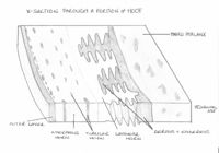
The epidermis of the horny hoof is termed the coronary epidermis. Its surface has a plantar aspect and newly formed hoof grows distally towards the plantar surface of the structure. Hoof wall is generally 5 - 10 mm in thickness and consists of 3 layers:
- A thin outer layer or "wall" of shiny, dense horn, which effectively seals the hoof against dehydration and penetration.
- A thicker, intermediate layer of amorphorous horn, interspersed with tubular horn which provides reinforcement. This layer makes up the bulk of the hoof.
- An inner, lamellar layer, where epidermis and dermis interdigitate, anchoring the dead portion of the hoof to the living surface of the third phalanx.
The pigmentation in the outer layer of hooves is derived from melanocytes in the coronary epidermis. Deeper layers contain little melanin and therefore appear lightly coloured or white. The unpigmented layer of keratin forms a white line on the sole of the hoof, which delineates the sole from the wall when the solar surface is viewed.
The keratin in the sole is formed by the epidermis of the plantar aspect of the third phalanx and reaches a maximum thickness of 1 cm. It is less resistant to abrasion than that of the outer layer of the hoof. The central frog of the sole of a horse is located between the sharply inflected bars of the wall and is softer and less rigid than the keratin of the wall of the hoof. Its elasticity is particulary important in the horse as pressure on the frog changes the angle of the walls when the horse stands on that foot. The change in angle, termed the hoof mechanism increases the blood circulation in the hoof, increasing the supply of nutrients to the coronary epidermis. This is closely correlated to hoof wall quality.
A band of soft, pliable horn termed the periople (analagous with the cuticle of human nails) lies over the proximal outer surface of the wall. This band widens at the back of the hoof to also cover the bulbs of the heels and part of the frog.
For further detail see the equine phalanges page.
Ruminant Hoof
Although the hooves of these species resemble the equine hoof, they differ in several ways:
- There are two separate main digits, compared with the single hoof of equids
- The wall is bent to form a dorsal border
- The bulb of the heel covers the entire caudal surface of the hoof and most of the plantar surface, leaving a small area of sole visible
- The interdigitating lamellae are smaller and less well developed.
The hooves of the main digits curve medially towards each other. The lateral digit carries more weight than the medial digit, and is larger. On the abaxial wall, the distal border makes contact with the ground along its entire length, whereas, on the axial wall, only does so toward the toe. The thickness of the wall increases towards the apex and the plantar surface.
The horn of the hoof generally grows at a rate of 5 mm per month, and in cattle allowed to move freely, growth should equal wear. In intensively kept cattle, growth exceeds wear, and foot trimming is required to maintain optimal shape and angle. The optimal angle of the toe from the ground is 50 degrees.
Dewclaws are present in most ruminants but do not make contact with the ground. They consist of wall and bulb and have no practical importance.
For further detail see the bovine lower limb page.
Porcine Hoof
The hooves of pigs are principally similar to those of ruminants, however the wall is straight, not bent medially at the toe, and have a soft bulb that is well distanced from the wall and sole. The hooves of the accessory digits are of the same structure as the principal digits, but only bear weight on soft ground.
Hoof trimming in pigs is rarely required due to the short lifespan of the farmed pig.
| Hoof - Anatomy & Physiology Learning Resources | |
|---|---|
 Test your knowledge using flashcard type questions |
Hoof Flashcards |
 Full text articles available from CAB Abstract (CABI log in required) |
The growth and adaptive capabilities of the hoof wall and sole: functional changes in response to stress. Bowker, R. M.; American Association of Equine Practitioners (AAEP), Lexington, USA, Proceedings of the 49th Annual Convention of the American Association of Equine Practitioners, New Orleans, Louisiana, USA, 21-25 November 2003, 2003, pp 146-168, 32 ref. Contrasting structural morphologies of "good" and "bad" footed horses. Bowker, R. M.; American Association of Equine Practitioners (AAEP), Lexington, USA, Proceedings of the 49th Annual Convention of the American Association of Equine Practitioners, New Orleans, Louisiana, USA, 21-25 November 2003, 2003, pp 186-209, 73 ref. |

