Small Animal Soft Tissue Surgery Q&A 14
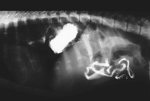
An eight-month-old, male English Bulldog is presented with a history of hypersalivation and regurgitation. On physical examination, mild dyspnea is noted. A contrast esophogram is performed and a lateral view radiograph is shown.
| Question | Answer | Article | |
| What is the diagnosis and what are the different types or categorizations of this abnormality? | Hiatal hernia. These hernias are classified as congenital or acquired (in animals most are congenital), and as a type I sliding hernia or type II periesophageal hernia (in animals most are type I). With type I hernias the phrenicoesophageal ligament is stretched, allowing the gastroesophageal junction to herniate back and forth into the thorax. With type II hernias the gastroesophageal junction remains stationery and the gastric fundus herniates through the esophageal hiatus alongside the esophagus. |
[[|Link to Article]] | |
| What is the surgical treatment for this problem? | This condition is rare in animals and the best surgical approach remains controversial. Successful treatment of three animals with hiatal hernia using a combination of three surgical techniques has been described. A modified Nissen fundoplication is performed to reduce gastroesophageal reflux, in conjunction with suture reduction of the esophageal hiatus and placement of a left fundic tube-gastropexy. The gastrostomy tube provides the additional advantages of allowing nutritional support, bypass of the esophagus and surgery site, and facilitates decompression of the stomach in the early postoperative period. Gas distension, presumably from an inability to belch, can cause discomfort after surgery. |
[[|Link to Article]] | |
| What is the prognosis? | The prognosis for complete relief of clinical signs is guarded. Review of reported cases shows approximately 25% success, and a mortality rate of 64%. |
[[|Link to Article]] | |
