| This article is still under construction. |
|
|
- Dermatophytes in dermatophytosis
- Caused by dermatophytes
- Microsporum - zoophilic
- Trichophyton - geophilic
- Epidermophyton - anthropophilic
- Common in many species, especially cats
- Hot, humid environment predisposes and viable fungi peripherally
- More common in young animals
- Produce proteolytic enzymes to penetrate surface lipid
- Fungal hyphae invade keratin -> break into arthrospores
- Epidermal hyperplasia (hyperkeratosis, parakeratosis, acanthosis) and inflammation
- Superficial perivascular dermatitis -> exocytosis (migration through epidermal layers) -> intracorneal microabscesses
- Exocytosis -> folliculitis -> furunculosis
- Highly variable lesions
- Normal -> eruptive nodular -> pseudomycetoma -> onychomycosis
- Grossly:
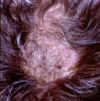
- Circular or irregular lesion, may coalesce
- Scaly to crusty patches
- Alopecia due to broken hair shafts and hairs lost from inflammed follicles
- Follicular papules and pustules
- Peripheral red ring (ringworm) due to dead fungi in areas of inflammation at centre of lesions and viable fungi peripherally
- Microscopically:
- Perifolliculitis, folliculitis or furunculosis
- Epidermal hyperplasia
- Intracorneal microabscesses
- Septate hyphae or spores may be found in stratum corneum and keratin of hair follicles


