Thrombocytes
Commonly known as platelets
Introduction
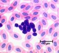
Thrombocytes are small anuclear fragments of megakaryocytes. They are membrane bound portions of the megakaryocyte cytoplasm and have a finely granular cytoplasm; they are much smaller than other blood cells at 2-3µm and have a lifespan of around 10 days in the circulation.
Development
Megakaryocytes develop from CFU-GEMM and from these thrombocytes are formed during thrombopoiesis.
Structure
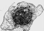
The thrombocyte is structurally divided into four zones:
- Peripheral zone is the outer membrane with a coat of glycocalyx
- Structural zone contains microtubules and actin and myosin filaments
- Organelle zone contains organelles such as mitochondria along with three different granule types
- Membrane zone is made of two types of membrane. One, the open canicular system, is membrane not used when the platelet budded off the megakaryocyte. The other, the dense tubular system, is membrane from the rough endoplasmic reticulum and is involved in calcium ion storage and regulation.
See reptile thrombocytes
Granules
- α granules are the most common and contain coagulation factors, fibrinogen, plasminogen, plasminogen activator factor and platelet derived growth factor (PDGF)
- δ granules contain ADP, ATP, serotonin and histamine and aid platelet adhesion and vasoconstriction
- λ granules contain lysosomes with hydrolytic enzymes to aid clot resorption
Actions
Thrombocytes play a number of roles in haemostasis. They adhere to exposed connective tissue in the walls of blood vessels forming platelet plugs, while releasing a number of factors from their granules. The glycocalyx on their surface provides a surface for fibrinogen to convert to fibrin leading to the formation of the secondary haemostatic plug. The platelets then contract (see structural zone) reducing the clot size. Finally lytic enzymes are released to break the clot down.
If platelet numbers fall below 50x109/l an animal is likely to haemorrhage after trauma and if the count falls below 30x109/l then spontaneous haemorrhage is a risk.
PDGF from the granules stimulates tissue repair in blood vessels by stimulating smooth muscle cells and fibroblasts.


