Tick Morphology
Jump to navigation
Jump to search
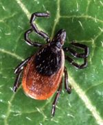
Hard ticks
- Hard, chitinous covering over dorsal surface called the scutum
- Unique to hard ticks
- Males have a scutum which covers the entire body surface
- Females have a scutum which only covers a small area behind the head
- Prominent biting mouthparts
- Festoons ('pie crust edging') around the posterior body margins
- Enamel coloured patches on scutum are present on ornate ticks
- Female hard ticks may swell up to 3 times their normal size when taking a blood meal
Soft ticks
- No scutum
- Mouthparts are not visible from dorsal surface
- Feed little and often as cannot swell as much as hard ticks
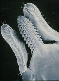
Mouthparts
- Sensory organs for locating a feeding site called palps
- Chelicerae for puncturing the skin
- The hypostome pushes through the wound made by the chelicerae where backwards pointing teeth lock the mouthparts into the skin
- Dorsal groove in the hypostome permits the flow of tick saliva and host blood
Feeding
- Ticks stand upright
- Chelicerae cut through skin creating a pool of blood
- Hypostome is inserted deep into the skin
- Mouthparts are cemented into place
- Ticks feed continuously
- Tick saliva flows into host and contains
- Histamine blocking agents to minimise the host inflammatory response
- Anticoagulants to ensure the free flow of blood
- Cytolysins to enlarge the feeding lesion
- Vasoactive mediators, enterases and carbohydrate splitting enzymes to increase the vascular permeability, facilitating feeding
- Paralytic toxins
- Host tissue is broken down leaving a zone of necrosis creating a feeding lesion


