Peripheral Nervous System - Histology
Introduction
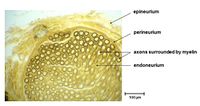
Nerves of the peripheral nervous system (PNS) are composed of numerous bundles of nerve fibers that are surrounded by connective tissue. This connective tissue also contains a specific layer that is specialised to neurons; the peri-neurium. The outer layer of this connective tissue is called the epineurium; which surrounds both the perineurium and the nerve itself. Individual neurons found within each bundle are surrounded by the endoneurium.
Each nerve fibre that is visible on histologic section represents an axon which is surrounded by the neurilemma or as it is also known, the sheath of a Schwann cell. However, depending on whether the cell is myelinated or unmyelinated, this layer may not be visible. Where present, the Schwann cell will be visible as as a dense layer immediately adjacent to the nerve fibre. This layer is then also immediately surrounded by the cytoplasm of the Schwann cell. The sheath and cytoplasm of the Schwann cell collectively form the neurilemma. Unmyelinated axons can often be seen running within small grooves of Schwann cells.
The information included on the page below is specifically to provide an insight into the histologic appearance of nerves within the PNS. Therefore knowledge of the terminology related to the anatomy and physiology of nerves is assumed and the PNS anatomy and physiology page should be reviewed to improve the understanding of the information provided below.
Histology
Myelinated Nerves
This image shows a cross-sectional histology of a myelinated nerve fibre. The section clearly shows numerous bundles of nerve fibres which are slightly out of focus, but appear as the globular contents of the nerve.
The nerve as a whole is surrounded by a dense layer of connective tissue; the epineurium. This layer often contains visible blood vessels and it is likely that there are two such vessels found to the left and upper-left of the nerve. Fat cells (adipose tissue) are also found around the epineurium, although none are clearly visible in this image.
Histology of Myelinated Nerve with lipid stain
The myelin stained here gives the nerve it's shape and support.
The perineurium and endoneurium provide a constant environment for the nerve fibres.
Histology of Myelinated Nerve (longitudinal section)
Histology of Ganglion
Histology of Autonomic Ganglion
Histology of Dorsal Root Ganglion
Comparison of the histology of the ganglion
| Autonomic Ganglion | Dorsal Root Ganglion |
|---|---|
| Multipolar cells | Pseudo-unipolar cells |
| Satellite cell layer incomplete.
(amphicytes) |
Satellite cells form complete covering.
(amphicytes) |
| Satellite cell layer incomplete.
(amphicytes) |
Satellite cells form complete covering.
(amphicytes) |
| Cells and fibre tracts interspersed. | Cells and fibre tracts in separate regions. |
| Nucleus usually off centre. | Nucleus centrally placed. |
| This article is still under construction. |


