Peripheral Nervous System - Histology
Introduction
Nerves of the peripheral nervous system (PNS) are composed of numerous bundles of nerve fibers that are surrounded by connective tissue. This connective tissue also contains a specific layer that is specialised to neurons; the peri-neurium. The outer layer of this connective tissue is called the epineurium; which surrounds both the perineurium and the nerve itself. Individual neurons found within each bundle are surrounded by the endoneurium.
Each nerve fibre that is visible on histologic section represents an axon which is surrounded by the neurilemma or as it is also known, the sheath of a Schwann cell. However, depending on whether the cell is myelinated or unmyelinated, this layer may not be visible. Where present, the Schwann cell will be visible as as a dense layer immediately adjacent to the nerve fibre. This layer is then also immediately surrounded by the cytoplasm of the Schwann cell. The sheath and cytoplasm of the Schwann cell collectively form the neurilemma. Unmyelinated axons can often be seen running within small grooves of Schwann cells.
The information included on the page below is specifically to provide an insight into the histologic appearance of nerves within the PNS. Therefore knowledge of the terminology related to the anatomy and physiology of nerves is assumed and the PNS anatomy and physiology page should be reviewed to improve the understanding of the information provided below.
Histology
Myelinated Nerves Cross-section
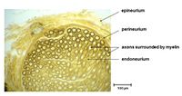
This image shows a cross-sectional histology of a myelinated nerve fibre. The section clearly shows numerous bundles of nerve fibres which are slightly out of focus, but appear as the globular contents of the nerve.
The nerve as a whole is surrounded by a dense layer of connective tissue; the epineurium. This layer often contains visible blood vessels and it is likely that there are two such vessels found to the left and upper-left of the nerve. Fat cells (adipose tissue) are also found around the epineurium, although none are clearly visible in this image.
Myelinated Nerve Cross-section (with lipid stain)
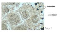
A lipid stain highlights aspects within the histology that contain lipid. In this case the main lipid bearing tissue within a nerve is the fat contained within the myelin sheath, or Schwann cells. In comparison with the image above, this image is taken at a higher magnification and shows the bundles of nerve fibres in more detail.
The epineurium is less visible in this image due to the higher magnification but is effectively around the upper-right of the image. Several blood vessels can be seen within the connective tissue around the bundles (seen as small holes within the connective tissue). As was discussed in the image above, the epineurium is often surrounded by adipose tissue. Due to the type of stain used for this image, several adipocytes can be seen to the right of the image. Each nerve bundle is surrounded by the perineurium and this can be clearly seen surrounding each.
Given the lipid stain, it is possible to distinguish the myelinated nerves from the unmyelinated nerves within each bundle. Therefore a large proportion of the nerves are myelinated within each bundle. These myleinated nerves appear at this magnification with a charactristic donut shape which represents the axon in the centre (which contains no lipid) and the surrounding Scwann cell (containing lipid).
Myelinated Nerve Longitudinal section
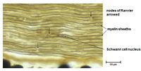
This image provides a longitudinal view of the edge of a nerve bundle. However, the image is not of sufficient quality to display a clear differentiation between the epineurium and perineurium. The epineurium is visible as the lighter strip at the base of the image.
Within the nerve bundle itself, the individual nerve fibres have a characteristic 'wavy' appearance. There are also a number of nuclei visible within the image and these are nuclei belonging to Schwann cells or to cells found within the endoneurium. At this magnification it is possible to identify some elements of the nerves themselves. In some of the fibres it is possible to visualise a faint central tubular structure which represents the axon of the nerve. (One is visible in the 6th nerve down from the top of the image). The lighter brown element of each nerve represents the myelin sheath whilst the darker brown lines surrounding both side of each nerve represents the thin cytoplasm of the Schwann cell (neurilemma cell).
Another clearly visible element of the individual nerves is indicated on the image by black arrows. These arrows indicate positions of nodes of Ranvier and represents the point at which two Schwann cells meet along the length of the axon. The histologic appearance of a node is as a constriction of the neurilemma.
Autonomic Ganglion Cross-section
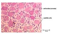
This cross-sectional image was stained with H&E and is at a sufficiently high magnification to identify very specific structures within the ganglion. The ganglion will contain a number of nerve fibres and cell bodies of the neurons. There are approximately 30 discrete nerve cell bodies visible in this image although there are no clear nerve fibres visible.
In some of the cell bodies it is possible to distinguish a faint ring which represents the centrally located axon. The cell bodies themselves are spherical and have retained a pale staining and in some there is an intensely stained nucleus. Surrounding these cell bodies are Satellite cells that appear to be continuous with the Schwann cells surrounding the nerve. These satellite cells often appear to be clustered and are much smaller than the cell bodies.
Dorsal Root Ganglion Cross-section
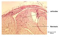
This cross-sectional view undertaken with H&E displays a number of characteristics of a dorsal root ganglion. The outer aspect of the ganglion shows the surrounding connective tissue of the epineurium within which a blood vessel is clearly visible. Outer adipose cells/tissue is also faintly visible to the top-right of the image.
Within the ganglion two distinct nerve bundles can be seen which are surrounded by the perineurium. The nerve bundle to the left of the image displays many similarities to that of the higher magnification cross-sectional image above (lipid stain image). At this magnification, individual nerves can be differentiated, although no specific details can be seen. The nerve bundle to the right contains similar nerve structures but also cell bodies and surrounding satellite cells that are very similar to the image previously reviewed above.
It should be noted that the areas of 'dead space' that are apparent within the nerve bundles are an artefact as a result of the histology process.
Comparison of the histology of the ganglion
| Autonomic Ganglion | Dorsal Root Ganglion |
|---|---|
| Multipolar cells | Pseudo-unipolar cells |
| Satellite cell layer incomplete.
(amphicytes) |
Satellite cells form complete covering.
(amphicytes) |
| Satellite cell layer incomplete.
(amphicytes) |
Satellite cells form complete covering.
(amphicytes) |
| Cells and fibre tracts interspersed. | Cells and fibre tracts in separate regions. |
| Nucleus usually off centre. | Nucleus centrally placed. |
| This article is still under construction. |
